Lumbosacral plexus dose with CT pelvimetry correlation in cervical cancer HDR brachytherapy
Candice Chin-Chin Yu,
Canada
PO-1791
Abstract
Lumbosacral plexus dose with CT pelvimetry correlation in cervical cancer HDR brachytherapy
Authors: Candice Chin-Chin Yu1, John Vincent Gan1
1Jose R. Reyes Memorial Medical Center, Radiotherapy, Manila, Philippines
Show Affiliations
Hide Affiliations
Purpose or Objective
Concurrent Chemoradiation is the established definitive treatment for patients with locally advanced cervical cancers. Recent developments in the utilization of intensity modulated radiotherapy technique (IMRT) and brachytherapy techniques such as image guided brachytherapy (IGBT) offers an advantage of increased target conformality. However, utilizing dose optimization may cause a potential problem in dumping doses to organs that are not routinely contoured such as the lumbosacral plexus (LSP) since they are not reflected in the dose volume histograms for plan evaluation. Our study investigates the LSP dose distribution with IGBT and correlating it with the patient’s pelvimetry characteristics.
Objectives:
1. To describe the brachytherapy dose distribution to the Lumbosacral plexus (LSP) among locally advanced cervical cancer patients treated with Image guided brachytherapy (IGBT).
2. To determine the correlation between pelvic anatomical parameters and brachytherapy dose received by the LSP.
Material and Methods
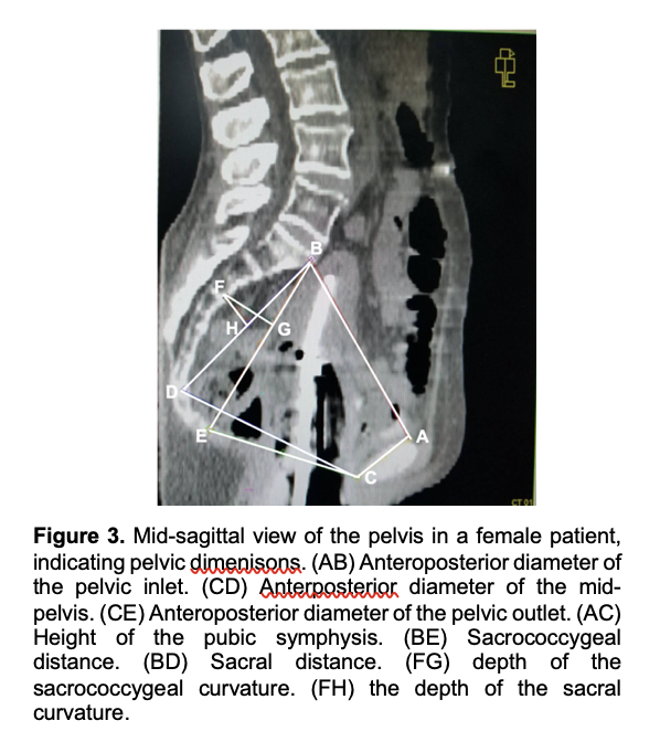
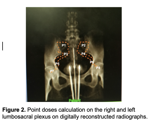
After meeting eligibility criteria, twenty nine patients with cervical cancer treated with conventional (cEBRT) and IGBT were included in the study. The LSP were contoured on the IGBT treatment plans and the mean volume, (maximum, minimum, and average) total doses to the whole LSP, point doses (P1, P2, P3, P4, P5, P6, P7, P8) were calculated. Pelvic dimensions were measured using three-dimensional reconstruction of computerized tomography (CT) images and were then correlated with LSP doses.
Results
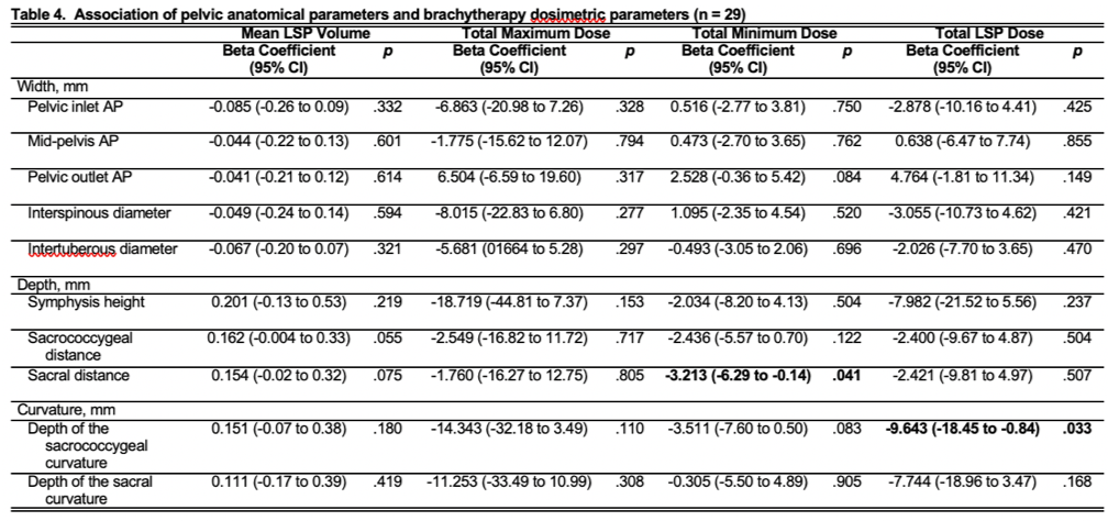
The average LSP volume was 10.1 ± 3.4 cc. The average total Dmax and total Dmin were 848.7 ± 271.3 Gy and 338.6 ± 62.2 Gy, respectively. The average total Dmean LSP dose 546.3 ±139.1 Gy. Point doses were highest at the levels of the inferior part of the sacroiliac joint (P3 and P4), followed by the ischial spine (P5 and P6), L5/S1 interspace (P1 and P2), and femoral neck (P7 and P8), respectively. There was evidence of significant but weak, inverse correlations between the sacral distance and Dmin (r = -0.3811), as well as between depth of the sacrococcygeal curvature and Dmean dose (r = -0.3969). Furthermore, there was evidence of significant but weak, inverse correlations between the intertuberous diameter and P5 (r = -0.4226) as well as P7 (r = -0.3871); the sacrococcygeal distance and P6 (r = -0.3704); as well the sacral distance and P2 (r = -0.4021), P6 (r = -0.3850), and P8 (r = -0.4213).
Conclusion
Conclusion: The delineation of LSP during IGBT planning may be helpful in accounting for LSP dose distribution in patients with small pelvic dimensions.