Radiomics predicts the location of local recurrence after reirradiation for head and neck carcinoma
PO-1633
Abstract
Radiomics predicts the location of local recurrence after reirradiation for head and neck carcinoma
Authors: Arnaud Beddok1,2,3, Valentin Calugaru1,3, Laurence Champion4,2, Christophe Nioche2, Gilles Crehange1,3, Irene Buvat2
1Institut Curie, Radiation Oncology, Paris, France; 2Institut Curie, Laboratory of Translational Imaging in Oncology (LITO). UMR (U1288). , Orsay, France; 3Institut Curie, Proton Therapy Center, Orsay, France; 4Institut Curie, Nuclear Medicine, Saint-Cloud, France
Show Affiliations
Hide Affiliations
Purpose or Objective
Curative reirradiation (reRT) is a promising alternative for the treatment of local recurrence (LR) of head and neck cancer (HNC). However, up to 50% of patients may experience a second LR within two years after the end of reRT. The aim of our study was to evaluate whether radiomics from FDG PET imaging could predict the localization of this second LR.
Material and Methods
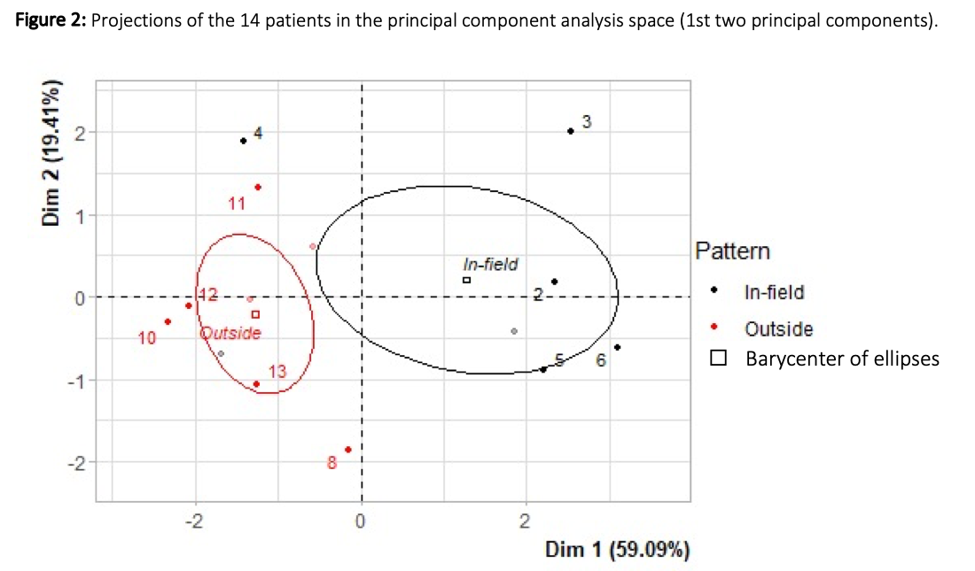
Among 23 patients re-irradiated with curative intent from 30/08/2012 to 08/04/2019 for advanced HNC, 14 patients had a second LR. For each of them, the reirradiated GTV was segmented based on the hypermetabolism on PET defined as SUV greater than 5. Twenty-nine parameters including three standardized uptake values (SUVs), five first order statistics derived from the gray-level histogram and 31 texture indices were extracted from these GTV using the LIFEX software after spatial resampling: 2 x 2 x 2 mm, intensity discretization: fixed bin size of 0.157, SUV units from 0 - 20. The volume of the second LR, called recurrent tumor volume (Vrecur, red line on Figure 1), was identified on PET scans. The LR were categorized as “in-field”, “marginal”, or “outside” if 100%, < 50% or < 20% of Vrecur was within the 95% isodose of the first recurrence GTV (green line on Figure 1), respectively. Student t.tests were used to compare the parameters extracted from both “in-field” and “outside” groups. Principal component analysis (PCA) and Ascending Hierarchical Classification (AHC) were performed to separate both groups. Correlograms were calculated to identify an association between the selected parameters.

Results
Among the 14 patients, seven had “in field” and seven had “outside” second LR, none was “marginal”. The value of two histogram indices (SUV_Kurtosis and SUV_std) and two texture parameters (GLCM_Correlation, GLCM_Contrast) were significantly different between both groups (p < 0.05). In PCA, the first two dimensions expressed 78.51% of the total dataset variance. “In-field” and “Outside” were the best separated variables on the plane (i.e. explained the best the distance between individuals, Wilks test p < 0.02, [figure 2]). “In-field” group corresponded to regions with high std and low Kurtosis, both features being associated with substantial signal heterogeneity. “Outside” group was associated with low GLCM_Correlation, std and GLCM_Contrast, suggesting rather homogeneous signal. The AHC analysis also found a cluster with high std, GLCM_Contrast, GLCM_Correlation, and low Kurtosis, corresponding to “In-field” patients. Kurtosis, std, GLCM_Contrast, GLCM_Correlation were not highly correlated (Pearson coefficient < 0.80).
Conclusion
This study suggests that before reRT, FDG PET radiomics features characterizing the signal heterogeneity in the GTV to be re-irradiated have different values in patients who will relapse “in-field” or “outside”, with “in-field” relapses associated with higher metabolic heterogeneity in the GTV than relapse occurring outside the GTV. A validation study is ongoing.