Evaluation of material properties retrieved with a radiotherapy specific CT scanner
PO-1628
Abstract
Evaluation of material properties retrieved with a radiotherapy specific CT scanner
Authors: Erik Pettersson1,4, Andreas Lindberg2, Ninni Drugge3, Anna Bäck3,5
1The Sahlgrenska University Hospital, Department of Therapeutic Radiation Phyiscs, Gothenburg, Sweden; 2The Sahlgrenska University Hospital, Department of Therapeutic Radiation Physics, Gothenburg, Sweden; 3The Sahlgrenska University Hospital, Department of Therapeutic Radiation Physics, Gothenburg, Sweden; 4Institute of Clinical Sciences, Sahlgrenska Academy at the University of Gothenburg, Department of Medical Radiation Sciences, Gothenburg, Sweden; 5Institute of Clinical Sciences, Sahlgrenska Academy at the University of Gothenburg, , Department of Medical Radiation Sciences, Gothenburg, Sweden
Show Affiliations
Hide Affiliations
Purpose or Objective
CT images are commonly
used for modelling of the patient for the absorbed dose calculation in external
beam radiotherapy. The dose calculation is sensitive to variations in density
and effective atomic number (EAN) of the tissue. The mass density (MD) and relative electron density (RED) images are usually
established from CT-numbers (Hounsfield units) using pre-defined CT-calibration
curves. New techniques are now available where information about material
properties can be provided directly from the CT software. This study compares
MD and RED obtained with single-energy CT (SECT) as well as RED and EAN obtained
with dual-energy CT (DECT) to theoretical reference values for materials with known
MDs and elemental compositions.
Material and Methods
The head sections of two electron density phantoms (Model 1467, Sun
Nuclear, SN) and (062M, CIRS) were placed inline in the holder belonging to the
first phantom (Figure 1). The SN phantom was setup with its tissue surrogate
rods, and a cylindrical container with 5 mg/cm3 iodine solution. The
CIRS phantom was setup with its tissue surrogates, as well as some non-tissue
equivalent materials (Table 1). The phantom setup was scanned with a
radiotherapy specific CT scanner (SOMATOM go.Open Pro, Siemens
Healthineers) with a SECT (120 kV) and a dual-spiral DECT (80 kV/Sn140 kV)
protocol. The SECT images were reconstructed with the Sd40 and Sm40 (DirectDensity™ (DD), Siemens Healthineers) kernels which provide RED and MD
images, respectively. The RED was also estimated from conventional SECT images
(Qr40 kernel) in a more traditional way
using a pre-defined CT-calibration curve created based on the CIRS phantom.
The DECT images were converted to RED and
EAN images in the Rho/Z application in (syngo.via.VB50A, Siemens
Healthineers). The values obtained with the different methods were
compared to the reference MD and theoretical reference RED and EAN of the
phantom inserts.
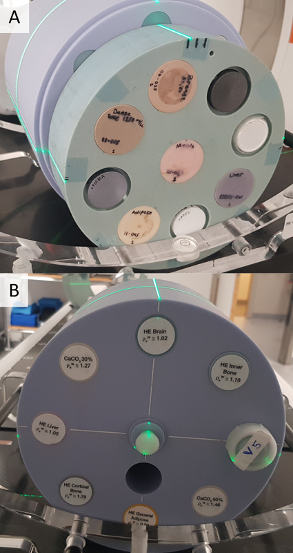
Figure 1. The head section of the CIRS 062M phantom is shown in A) and an
opposite view of the setup that shows the Sun Nuclear 1467 phantom is shown in B).
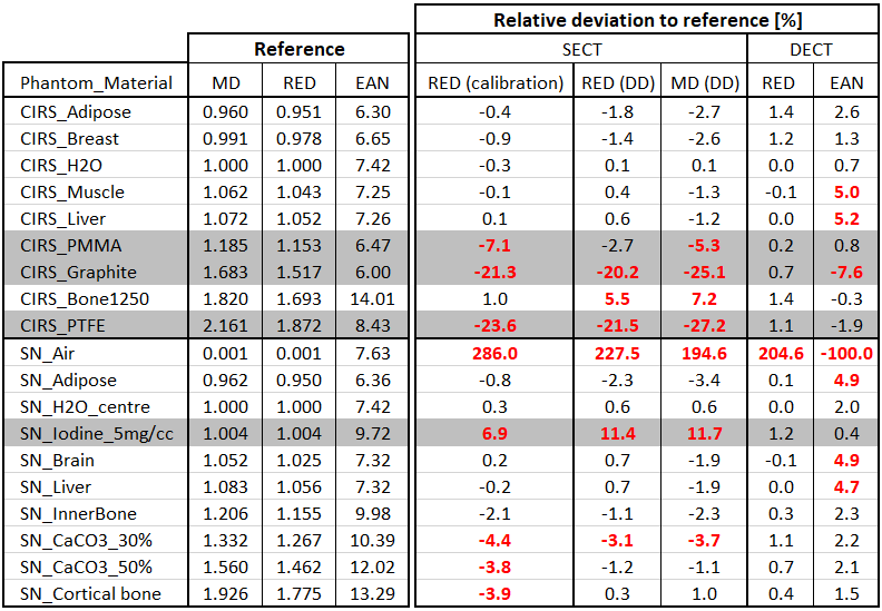
Table 1. The reference values of the MD, RED and EAN, as well as the
relative differences of the studied methods compared to the reference values.
Deviations >±3% are written in red. Non-tissue equivalent materials are
shaded in grey.
Results
Both SECT methods
underestimated the RED and MD for PMMA, graphite and PTFE (Table 1). The RED
and MDs of the iodine solution were overestimated for the SECT methods,
although less using the traditional calibration compared to DD. However, the traditional
CT-calibration resulted in an underestimation of the RED of the three most
compact bone surrogates in the SN phantom with 4%. DECT provided RED values
within 1.4% for all materials in the phantom setup. Larger deviations were
observed for the EAN.
Conclusion
Generally, the DECT method resulted in RED values
closer to the references values compared to both SECT methods studied and
especially for non-tissue-equivalent materials.