Peripheral dose sparing of volume-of-interest CBCT using a novel 2.5 MV low-Z beam
Jennifer Borsavage,
Canada
PO-1627
Abstract
Peripheral dose sparing of volume-of-interest CBCT using a novel 2.5 MV low-Z beam
Authors: Jennifer Borsavage1, Amanda Cherpak2, James Robar2
1Dalhousie University, Department of Physics and Atmospheric Science, Halifax, Canada; 2Dalhousie University, Department of Radiation Oncology, Halifax, Canada
Show Affiliations
Hide Affiliations
Purpose or Objective
Megavoltage imaging offers the advantageous perspective from the treatment beam's-eye-view (BEV), however, due to the high energy photons in an MV spectrum, images acquired with this modality suffer from limited image quality per unit imaging dose. A commercial 2.5 MV beam at our centre was recently modified to replace the copper target with a low-Z sintered diamond alternative. This 2.5 MV beam contains a significant population of diagnostic-energy photons, yielding improved planar CNR versus dose characteristics compared to the commercial imaging beam. To further reduce the dose to healthy tissues in 2.5 MV low-Z CBCT, the MLC was used to collimate the beam to the relevant anatomy, through a volume-of-interest (VOI) CBCT approach in this study.
Material and Methods
A sintered diamond target was integrated into the TrueBeam (Varian Medical Systems, Palo Alto, CA) target arm replacing the commercial copper imaging target. Using this experimental beam, the sinuses of an anthropomorphic phantom was imaged to investigate the dose sparing capacity of the VOI technique. VOI regions were defined in the treatment planning system as concentric volumes where the smallest VOI conformed to the sinuses, and additional VOIs were created by adding 0.5, 1, and 1.5 cm margins (Figure 1). MLCs were fit to the BEV of each volume throughout a full 360 degree arc. The corresponding MLC sequences were used to construct XML scripts for acquisitions using Developer Mode. For comparison, an additional full field-of-view (FOV) CBCT was also acquired. During each CBCT acquisition, calibrated EBT3 radiochromic film was placed between phantom slices along the transverse plane to measure axial dose distributions.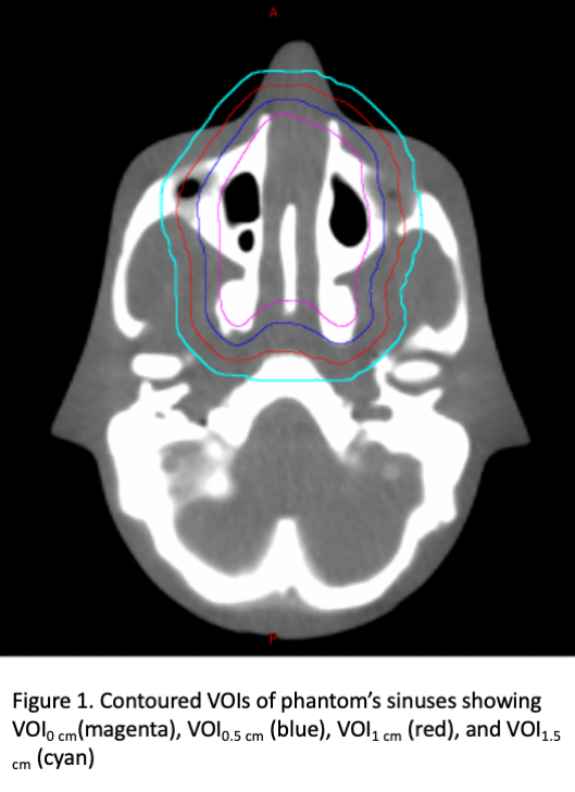
Results
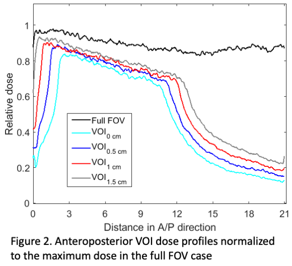
Compared to the full FOV acquisition, a decrease in imaging dose was observed for each of the four VOI cases in the anteroposterior direction (see Figure 2). Imaging dose decreased with VOI dimension, with dose reductions within the VOI ranging from 7% to 20%, compared to the full FOV case. Outside of the VOI, imaging doses decreased to 23%, 20%, 15% and 12% of the full FOV maximum dose.
Conclusion
This study demonstrates the dose sparing capabilities of 2.5 MV low-Z VOI CBCT. Compared to the full FOV acquisition, imaging dose within the VOI decreased by up to 20% by collimating the beam with the MLCs. This dose reduction results from decreased scatter in the treatment head and within the phantom compared to the full FOV case. While this study focused on VOI acquisition using a standard EPID, it is anticipated that imaging doses could be further reduced using an efficient multilayer imager in combination with this technique.