Image reconstruction using the PETITION PET scanner aimed at biologically guided proton therapy
Shubhangi Makkar,
Switzerland
PO-1602
Abstract
Image reconstruction using the PETITION PET scanner aimed at biologically guided proton therapy
Authors: Shubhangi Makkar1,2, Marina Béguin2, Günther Dissertori2, Judith Flock2, Cristian Fuentes2, Jan Gajewski3, Jan Hrbacek1, Keegan McNamara1,2, Christian Ritzer2, Antoni Rucinski4, Damien C. Weber1,5, Antony Lomax1,2, Carla Winterhalter6,2
1Paul Scherrer Institute, Center for Proton Therapy, Villigen PSI, Switzerland; 2ETH Zürich, Department of Physics, Zürich, Switzerland; 3Institute of Nuclear Physics, PAS, Krakow, Poland; 4Institute of Nuclear Physics, PAS, Krakow, Switzerland; 5University Hospital of Bern and Zürich, Radiation Oncology Department, Bern, Zürich, Switzerland; 6Paul Scherrer Institute, Center for Proton Therapy, Zürich, Switzerland
Show Affiliations
Hide Affiliations
Purpose or Objective
In the context of the PETITION project, a PET scanner is being developed for range verification and online hypoxia guided proton therapy for gantry 2 at PSI. It is designed as a table mounted system with an opening for proton irradiation and can rotate to allow treatment from multiple directions. The scanner opening causes some projections to be lost and gives rise to artefacts. This simulation-based work investigates the correction of this artefact in the image reconstruction process by using the information from four scanner positions simultaneously.
Material and Methods
The PETITION scanner was modelled in the GATE Monte Carlo toolkit (v8.2). A water cylinder (H = 4 cm, ∅ = 2 cm) filled with 1 MBq of back-to-back gamma activity was simulated at the center of the Field of View (FOV) of the scanner for 40 seconds. The coincidences were sorted with a timing window of 4 ns and reconstructed using CASToR (v3.1). To account for the open ring design of the PETITION scanner, a new look up table was generated, and the crystal numbers were matched in GATE and CASToR. Ordered Subset Expectation Maximisation (OSEM) algorithm was used for reconstruction with 4 subsets and 12 iterations. In the first workflow, one scanner position was considered, whereas in the second workflow, the data from four scanner positions were taken into account simultaneously by adding up the reconstructed data in the image space. Furthermore, to validate the obtained images, the same cylindrical phantom was also simulated and reconstructed with a full ring scanner design.
Results
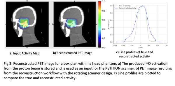
The Structural Similarity Index Measure (SSIM) and Mean Squared Error (MSE) have been computed between the normalised reconstructed images obtained by the full ring scanner and the PETITION open ring design. Image quality is substantially degraded when using one scanner position only (Fig 1a, SSIM of 0.88 and MSE of 0.12). When taking four positions of the rotating scanner into account (indicated schematically in Fig 1b), image quality can be restored to a SSIM of 0.97 and MSE of 0.01. The same reconstruction workflow has been applied to reconstruct the activation caused by irradiation with a proton beam in a head phantom, resulting in good agreement with the expected image (Fig 2c).
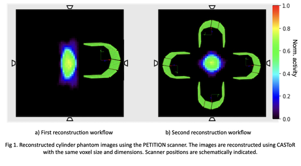
Conclusion
The reconstruction workflow has been setup for the novel, open ring PETITION scanner. The reconstructed image (Fig 1) using the approach of rotating the scanner confirms the validity and demonstrates that the reconstructed image is equivalent to the image obtained by the full ring design. The final image has been substantially improved by considering multiple scanner positions simultaneously. Next steps will investigate a machine learning approach to the reconstruction process.