Evaluation of synthetic CTs generated from T2-weighted MRIs of prostate cancer patients
Sebastian Andersson,
Sweden
PO-1597
Abstract
Evaluation of synthetic CTs generated from T2-weighted MRIs of prostate cancer patients
Authors: Sebastian Andersson1, Isabell Steinseifer2
1RaySearch Laboratories, Research, Stockholm, Sweden; 2Isala, Department of Radiation Oncology, Zwolle, The Netherlands
Show Affiliations
Hide Affiliations
Purpose or Objective
In radiotherapy, MR imaging is used
for delineation in an increasing degree due to its superior soft tissue
contrast. However, CT images are still needed for treatment planning, as MR
images lack tissue density information. Accurate MR-to-CT synthesis is a
crucial step towards an MR-only workflow in radiotherapy. By removing the need
of CT imaging, a clinic can both save time and get rid of potential MR-CT
registration uncertainties. This work
evaluates an algorithm for synthetic CT (sCT) generation, available in the
research version of RayStation 10A (RaySearch Laboratories, Stockholm, Sweden).
Material and Methods
This study included T2-weighted TSE
MRI (VISTA) (Philips Healthcare, Best, The Netherlands) and CT Big Bore
(Philips) pelvic images of 55 patients in treatment position. The MR and CT images
were registered deformably to reduce the anatomical differences. The synthetic CTs
were generated in a semi-supervised fashion, using a CNN architecture similar
to CycleGAN but including an extra paired term to account for the existence of paired
data in this context. 35 patients were used for training of the sCT model and 20
patients were used for evaluation of the resulting sCT images.
Mean absolute errors (MAEs) were calculated
between the Hounsfield units (HU) of the deformed CT and the sCT for each
patient. The original VMAT treatment plans were recalculated on both the
deformed CT and the sCT. The dose differences between the CT based and the sCT
based dose distributions were calculated for D1, D2, D50, D95, D98, D99 and
average dose, for the following ROIs: CTV, PTV, rectum, anal canal, bladder,
left femoral head and right femoral head.
A research version of the commercial
treatment planning system RayStation 10A was used for both image conversion and
dose calculation.
Results
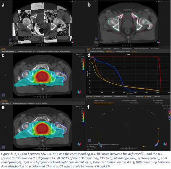
The mean MAE between the deformed CT
and the sCT was 41.5 ± 9.6 HU. Figure 1a
shows the fusion between the MRI and the corresponding sCT, with a very good conformity
of the structures. Figure 1b shows a fusion of the deformed CT and the sCT with
minor differences. Furthermore, the dose distribution on the deformed CT and
the sCT of one patient (c+e), as well as the DVH’s (d) and a dose difference
map (f). The DVH’s overlay each other, no difference between the deformed CT
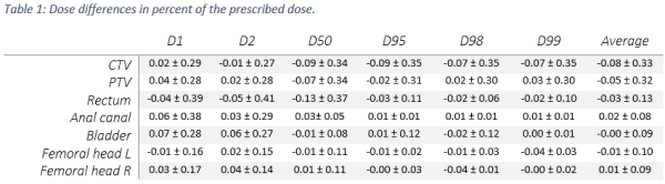
and the sCT can be seen. In Table 1 the mean dose difference of the
prescribed dose is shown in percent for all dose statistics. The
dose comparison shows a very good agreement with the deformed CT, the mean dose
differences were close to 0% and no standard deviation above 0.5%.
Conclusion
Using the synthetic CT images generated from T2-weighted MR images results
in clinically insignificant dose differences compared to dose calculated on the
deformed CT. Therefore, sCT images generated with this algorithm would allow
for a MR-only workflow in radiotherapy planning.