A simple proton-radiography system for accurate RSP measurements in proton therapy
Francesco Olivari,
The Netherlands
PO-1591
Abstract
A simple proton-radiography system for accurate RSP measurements in proton therapy
Authors: Francesco Olivari1, Emiel van der Graaf1, Marc-Jan van Goethem1, Sytze Brandenburg1
1University Medical Center Groningen (UMCG), CRCG, Groningen, The Netherlands
Show Affiliations
Hide Affiliations
Purpose or Objective
Proton radiography (pRG) can be used to
optimize proton relative stopping power (RSP) predictions from
X-ray-imaging-based techniques to improve the quality of proton therapy
treatment plans. In this work, we present a simulation study for a simple
pRG-system with the potential of being routinely used in the clinic and with
sufficient RSP accuracy to be used for improving X-ray-based RSP predictions.
Material and Methods
The simulated pRG-system consists of a thin and
finely 2D-pixelated detector measuring the energy deposited by primary protons
(protons produced at the source) and their fluence distribution. The mean
energy deposited per primary proton is converted into residual range in water
with a calibration function. By irradiating the detector without and with a
sample placed before the screen the RSP of the material is obtained from the
residual range difference and sample thickness.
The RSPs of 12 different materials (Gammex
human-tissue-equivalent materials, plastics, metals, and carbon) have been
determined from Monte Carlo simulations. To estimate the accuracy of our method
in the simulation framework, the RSPs obtained with the screen are compared
with reference RSPs obtained from simulations of irradiations of a water tank in
which the residual range of protons is determined: the RSP of a material is
derived from the range shift with a sample before the tank and the sample thickness.
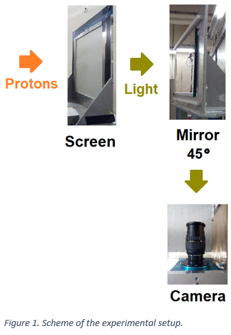
The system is realized experimentally with a
scintillator screen coupled with a CCD camera (Figure 1): the light yield,
which is proportional to the energy deposit in the screen, is measured. The
mean light yield per incident primary proton is calculated using the data of
the fluence distribution scored with simulations as a proxy of the real fluence
distribution, and it is converted into residual range in water. For the
materials considered in simulations, the experimental RSPs are determined with
the screen and compared with reference RSPs derived from residual range
measurements in a water tank.
Results
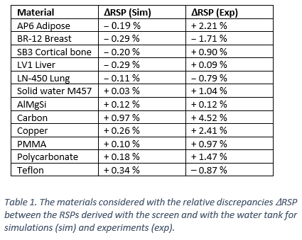
The
simulations show an agreement within 1% of the RSPs derived from the screen and
those from the water tank for all materials. The experimental results show an agreement
within 1% for 6 materials (3 are human-tissue-equivalent), while the agreement
is better than 5% for all materials (Table 1). The agreements are similar with the
results from Monte Carlo simulations and experiments of [1]. The RSPs of 5 of
the Gammex materials were also derived in [2,3]: our results agree with theirs
within from 1% to 6%, except for lung (discrepancies around 10%).
Conclusion
The method proposed seems to have the potential
to provide RSP predictions with an accuracy better than 1%: in the simulations
done, this was possible for all materials considered; in the experiments, for
half of the materials.
[1] doi.org/10.1016/j.nimb.2018.09.015
[2] doi.org/10.1088/1361-6560/ab9981
[3] doi.org/10.1088/0031-9155/61/22/8085