Using novel silica bead TL dosimeters to determine output factors for a kV radiotherapy unit
John Kearton,
United Kingdom
PO-1565
Abstract
Using novel silica bead TL dosimeters to determine output factors for a kV radiotherapy unit
Authors: John Kearton1, Antony Palmer1, Vasileios Goudousis2, Shakardokht Jafari1
1Portsmouth Hospitals University NHS Trust, Medical Physics, Portsmouth, United Kingdom; 2University of Surrey, Department of Physics, Faculty of Engineering and Physical Sciences, Guildford, United Kingdom
Show Affiliations
Hide Affiliations
Purpose or Objective
It is common practice to use generic data published in BJR supplement 25 [BJR 1996] to derive kV radiotherapy treatment output factors (MU per cGy).
Due to difficulties in measuring dose for small fields at low energies this
is an acceptable data sourse. However, the data is based on HVL and takes no
account of the beam characteristics of specific kV treatment units which can
lead to uncertainty. The objective of
this work was to devise a novel method using small silica bead
TL detectors to accurately measure the output factors for kV radiotherapy
treatments, and compare results to a traditional ionisation chamber technique.
Material and Methods
Two independent dosimetry methods (silica TL beads, 1.1mm thick, and
CCO4 ionisation chamber, 0.04cm3
volume, 4 mm cavity diameter) were used to measure the dose delivered for a range
of treatment applicator field sizes (2-20cm diameter) and lead cut-outs, for
the Xstrahl D3300 treatment unit, over a range of energies (70-250kV).
The novel aspect of this work was to experimentally determine output
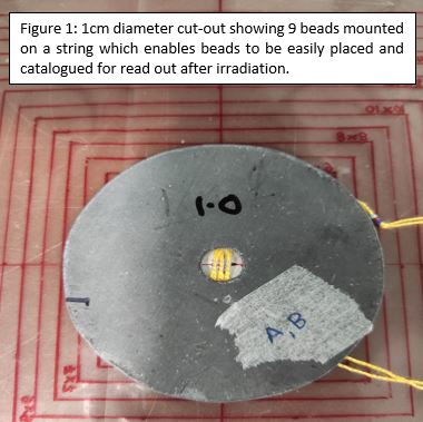
factors using silica beads, which have only had limited use in kV energies [Palmer et al 2017] and [Jafari et al 2014]. Due to their small size, nine beads were used for each dose
measurement to reduce uncertainly. Beads were placed on the surface of a block
of solid water with a stand-off of 6 mm,
see Figure 1.
Calibration of the glass beads was performed for each energy against a
traceably calibrated ionisation chamber to generate an energy specific
calibration factor. This was in addition to bead-specific individual
sensitivity calibration using 6 MV photons in a reference condition factors,
completed due to small variation measured across a batch of beads. The glass
bead TL signal was read-out using a TLD reader (Toledo 654).
Results
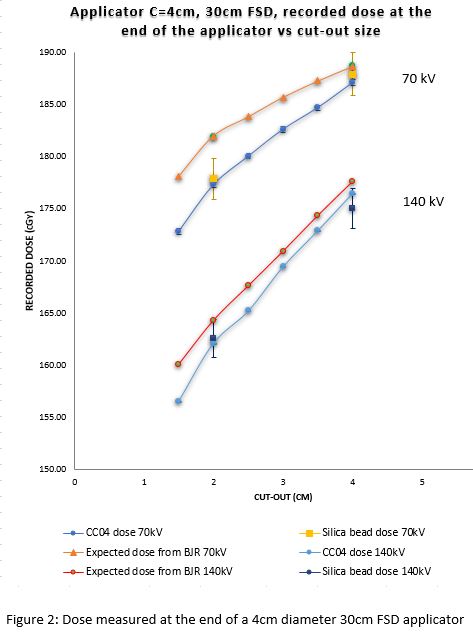
Differences were observed between the experimentally determined output
factors using the two detectors and existing plan data obtained from BJR 25, as
illustrated in Figure 2 for a 4cm applicator with various lead cut-outs The
experimental output factors measured using the CC04 ionization chamber and
silica beads TLDs were in agreement, within experimental uncertainty. These
discrepancies were higher for the smallest and largest field sizes, for all
energies. The beads and CC04 results differed by up to 1% (uncertainty +/-
1.1%), the mean value of which was up to 3.1% different to BJR data.


Conclusion
The output factors for a kV treatment unit have been successfully
measured using silica bead TL dosimeters, verified against an ionisation
chamber. Statistically significant difference was measured from the current
clinically used BJR 25 data and should be adopted clinically. This work also
shows that silica beads can be successfully used to verify the dose delivery of
small field sizes on a kV unit, important
as they are only 1.1mm thick (compared to 4mm diameter for the CC04) so can be
used to measure very small field sizes, down to 1cm diameter cut-outs in this
work.