Monte-Carlo modelling of Hp(10) in a superficial treatment room to inform radiation risk assessments
PO-1551
Abstract
Monte-Carlo modelling of Hp(10) in a superficial treatment room to inform radiation risk assessments
Authors: Paul O'Connor1, Margaret Moore2, John Cronin2
1National University of Ireland Galway, Medical Physics, Galway, Ireland; 2University Hospital Galway, Radiotherapy, Galway, Ireland
Show Affiliations
Hide Affiliations
Purpose or Objective
Current Irish radiation protection legislation S.I.30 requires hospitals to have radiation risk assessments that include detailed estimates of potential doses which could occur due to any unintended exposures. To quantify such radiation exposure, a Monte Carlo (MC) simulation model was created to calculate the Hp(10) value at any point in a superficial therapy room over the duration of a treatment. To verify that model, direct measurement of Hp(10) within that superficial therapy room was carried out.
Material and Methods
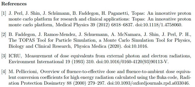
The MC model was created using Geant4-based MC toolkit, TOol for PArticle Simulation (TOPAS) [1][2] and the Hp(10) phantom was created using an ICRU slab as defined in ICRU report 47 [3], with a fluence scorer placed 10 mm below the surface of the phantom. This fluence was then converted into Hp(10) values using the conversion coefficients as set out by Pelliccioni [4]. Measurements were then taken at varying locations in the treatment room using an Electronic Personal Dosimeter (EPD) calibrated to measure Hp(10) values to verify the model.
Results
Firstly, the relationship between histories in the model and the monitor units on the machine was found to be H(100MU) = 1.63x10^(14) histories/100 MU for the 100 kV beam and H(100MU) = 1.82x10^(14) histories/100 MU for the 140 kV beam. Measurements of the Hp(10) value were then taken for 100 MU, with the detector measuring values up to 60 µSv for the 100 kV beam and up to 133 µSv for the 140 kV beam. These measurements were compared to the values given by the model, with the Hp(10) values predicted by the model being within 10% of the measured values for the 100 kV beam and within 15% of the measured values for the 140 kV beam. When the model was applied to a range of clinical scenarios, the Hp(10) value was calculated to be ranging from 4 mSv when the exposed person was 50 cm from the patients position to 16 mSv when the exposed person was 25cm from the patient position, for a typical treatment delivering 500 MU at 140 kV.
Conclusion
A MC simulation model of the superficial x-ray therapy unit was successfully created for the 100 kV and 140 kV beam energies. This model was verified by comparing it to measured values. The model can quantify radiation exposure for a variety of scenarios of potential unintended exposures and identify sources of highest risk. Risk assessments can be dynamically adapted over time using the model, to reflect any changes in the controlled area, as recommended by the EPA. The model represents a valuable tool for physicists and especially radiation protection advisers, as it will allow them to create more reliable and realistic radiation risk assessments.
