MRI-based Synthetic CT images for IMPT Treatment Planning of Nasopharyngeal Carcinoma Patients
PO-1503
Abstract
MRI-based Synthetic CT images for IMPT Treatment Planning of Nasopharyngeal Carcinoma Patients
Authors: Shupeng Chen1, Yinglin Peng2, Yimei Liu2, Chong Zhao2, Xiaowu Deng2, An Qin1, Di Yan1, Craig Stevens1, Rohan Deraniyagala1, Xuanfeng Ding1
1Beaumont Health, Radiation Oncology, Royal Oak, USA; 2Sun Yat-sen University Cancer Center, Radiation Oncology, Guangzhou, China
Show Affiliations
Hide Affiliations
Purpose or Objective
To investigate the feasibility of using an MRI-based synthetic CT image (SCT) generated via a generative adversarial network (GAN) for intensity-modulated proton therapy (IMPT) treatment planning of nasopharyngeal carcinoma (NPC) patients.
Material and Methods
T1-weighted MR images and paired CT (PCT) images were obtained from 158 NPC patients with radiotherapy immobilization. Deformable image registration was performed between each MR and PCT image for each patient to create an MR-CT pair. Thirteen pairs were randomly chosen as independent test sets and the remaining 145 pairs (10 for validation and 135 for training) were used to build a conditional GAN model, including a residual-Unet as a generator and a 6-layer convolution neural network as a discriminator. For each test patient, SCT was generated using the generator with the MR image as input. A 4-beam IMPT plan was created and optimized on the corresponding PCT, and the dose matrix was recalculated on the SCT. The dosimetric accuracy was evaluated by using the clinically relevant dose-volume histogram (DVH) parameters and 3D gamma index analysis.
Results
The mean absolute error between the PCT and SCT images were (89.64+20.54)HU within the body. Figure 1 shows the MR, PCT, SCT, and HU errors for a patient with an average performance of CT number accuracy. The DVH parameters discrepancy between dose matrices calculated on PCT and SCT were (0.13+0.13)%, (0.4+0.44)%, (0.81+0.78)%, (1.25+1.26)%, (1.24+0.78)%, and (1.35+1.1)% for CTV1-D95, CTV2-D95 (involved nodes), Left-parotid mean dose, right-parotid mean dose, brain stem D1, and spinal cord D1, respectively. Figure 2 shows the dose matrices calculated on the PCT and SCT as well as the DVH of critical structures and targets for a patient with an average performance of dosimetric accuracy. The 3%/3mm (10% threshold) gamma passing rate was (97.26+2.35)% within the head and neck region for the 13 test patients.
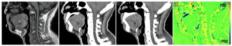
Figure 1 (Left to right) MR, paired CT (PCT) and synthetic CT (SCT) as well as the HU error for a patient with an average performance of CT number accuracy (mean absolute error = 86.66 HU).
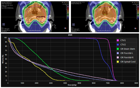
Figure 2 Dose matrices calculated on paired CT (PCT) (top left) and synthetic CT (SCT) (top right) as well as the dose-volume histogram (bottom) (solid line: PCT and dashed line: SCT) for a patient with average performance of dosimetry accuracy (3%/3mm gamma passing rate = 97.7%)
Conclusion
Overall, the SCT generated from MRI using GAN model achieved clinical acceptable dosimetric accuracy of IMPT planning for NPC patients. However, further study is needed to validate the SCT algorithm on a larger patient cohort before clinical implementations.