Copenhagen, Denmark
Onsite/Online

ESTRO 2022

Session Item

Inter-fraction motion management and offline adaptive radiotherapy
6029
Poster (digital)
Physics
Comparison of CBCT Hounsfield Unit to density conversion methods for treatment plan evaluation
Barbara Vanderstraeten, Belgium
PO-1493

Abstract

Comparison of CBCT Hounsfield Unit to density conversion methods for treatment plan evaluation
Authors:

Barbara Vanderstraeten1, Noémie Johnston2, Jorn Ketelaars2

1Ghent University, Ghent University Hospital, Radiotherapy-Oncology, Gent, Belgium; 2Ghent University, Human Structure and Repair, Gent, Belgium

Show Affiliations
Purpose or Objective

Daily CBCT images can be used to evaluate the need for treatment plan adaptation in head and neck cancer (HNC). To assess the impact of anatomical changes, CBCT dose calculation is required. CBCT images generally have a lower image quality and smaller field-of-view (FOV) compared to CT. We compared two CBCT Hounsfield Unit (HU) to mass density conversion methods for treatment plan evaluation.

Material and Methods

CT images were taken using a Toshiba Aquilion CT scanner at 120 kV, while CBCT images were collected using the on-board XVI system on an Elekta Synergy linear accelerator with a fast head and neck scanning protocol (200° rotation, 100 kV, small FOV and no filter).
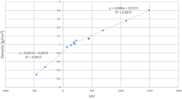
First, a site-specific conversion (SSC) curve for HNC was created by placing Gammex phantom inserts with known mass densities into a small custom-made phantom (14x14x11 cm³), resulting in a bi-linear SSC curve for HNC (figure). 

Secondly, a patient-specific conversion (PSC) method available in RayStation TPS was used, automatically attributing different ranges of HU values to 6 materials (air, lung, adipose tissue, cartilage/bone and other high-density material) with corresponding mass densities.

The SSC and PSC methods were evaluated on the anthropomorphic Alderson RANDO phantom as well as on 7 patient data sets. Only HNC patients with a re-planning CT (rCT) for treatment adaptation were retrospectively selected. Dose calculation was performed on the first re-planning CBCT (rCBCT).  All patients received VMAT with 6MV photons and a SIB up to 69.12 Gy in 32 fractions. Dose calculations were performed in RayStation v9 using the CC algorithm.

Dose comparisons between rCT, SSC-rCBCT and PSC-rCBCT were performed by means of DVHs, dose statistics and 3-D gamma (3%, 3mm) maps. PTV and OAR ROIs were delineated on rCT and transferred onto the rCBCT using a rigid registration between both image sets.

To evaluate the potential aid of both methods in clinical decision making, the clinical goals of the original treatment plan were evaluated on the CBCT’ images on which the decision to re-plan was based.

Results

For the RANDO phantom, the absolute differences in average mass density between SSC and PSC for different ROIs were < 0,1 g/cm³. The relative dose differences for D2, D50, Dmean and D95 were all < 0,5%. Gamma passing rates of SSC and PSC compared to the dose calculated on CT were 99,9% and 99,8%, respectively.

For the patients, the time between the original planning CT and CBCT’ ranged from 16 to 39 days, while the time between rCT and rCBCT ranged from 1 to 3 days. Gamma passing rates ranged between 92,7% and 99,2% for rCT-SSC and between 92,9% and 99,6% for rCT-PSC, respectively.

For support in clinical decision making towards re-planning, only the PTV showed changes in the achievement of the clinical goals.

Conclusion

A SSC HU to density conversion curve was created for HNC. Both SSC and PSC are suitable for CBCT dose assessment for treatment plan evaluation in the framework of adaptive radiotherapy.