Accuracy of inter-fraction patient positioning in Ocular Proton Therapy (OPT).
Martijn Hol,
The Netherlands
PO-1484
Abstract
Accuracy of inter-fraction patient positioning in Ocular Proton Therapy (OPT).
Authors: Martijn Hol1, Myra Rodrigues2, Yvonne Klaver3, Kees Spruijt4, Jasper Kouwenberg4, Eleftheria Astreinidou1, Coen Rasch1
1Leiden University Medical Centre, Radiotherapy, Leiden, The Netherlands; 2Holland Proton Therapy Center, Physicians, Delft, The Netherlands; 3Holland Proton Therapy Center, Physicians, Delft , The Netherlands; 4Holland Proton Therapy Center, Physics, Delft, The Netherlands
Show Affiliations
Hide Affiliations
Purpose or Objective
In this
study we quantify the accuracy of the day-to-day positioning in Ocular Proton
Therapy using fiducials.
Material and Methods
Treatment
of an ocular patient with proton therapy consists of five stages: fiducial
placement, simulation, treatment planning, dry run and treatment. Four Tantalum
fiducials (2.5 mm diameter, 0.17 mm thickness) are used to localize the tumor
perioperatively. During simulation, the patient is positioned in an upright
position in a robotic chair and fixated with a bite block, a thermoplastic mask
and strap to support the back of the head. The fiducials are located on two
orthogonal x-ray images. Localization parameters, i.e. clip-tumor distances,
fundus and ultrasound and MR images are used in the Eclipse Ocular Proton
Planning system (Varian Medical Systems, Inc.) to create the treatment plan.
Before treatment, the position verification is done by matching the reference fiducials
images of the treatment plan to the x-ray images. The patient’s position is
corrected by the robotic chair for translations only.
We investigated the day-to-day position
variations based on the Tantalum fiducials position in the x-ray images just
prior to the treatment. For this, analyses the four fiducials on the x-ray
images are localized using ImageJ. The
fiducial positions in the reference images created in the treatment planning
system were extracted from the dicom file via a self-developed Python script.
The day-to-day accuracy is calculated by comparing the center of gravity of the
fiducials position of the x-ray images to the center of gravity of the
fiducials found in the reference images. 60 consecutive patients were analyzed
with a total of 240 fractions. From the collected data the mean deviation,
random error and systematic error of the patient population are calculated.
Results
The
day-to-day statistics of the patient population are shown in table 1. As
expected the mean deviation is around zero millimeters. The largest deviation is
found in the longitudinal direction (AP-PA for the patient) both in mean
deviation, random and systematic error and is caused by tightening the strap at the back of the
patient’s head. This is done manually for each fraction. Figure 1 shows the
deviation in the patient positioning as a function of the percentage of the
found deviations. Also this graphs shows larger deviations in the longitudinal direction.
Table 1: Day-to-day statistics of OPT patient population
| Lateral | Longitudinal | Vertical |
| Mean deviation [mm] | -0.11 | -0.04 | 0.03 |
| Random error [mm] | 0.28 | 0.56 | 0.34 |
| Systematic error [mm] | 0.19 | 0.43 | 0.23 |
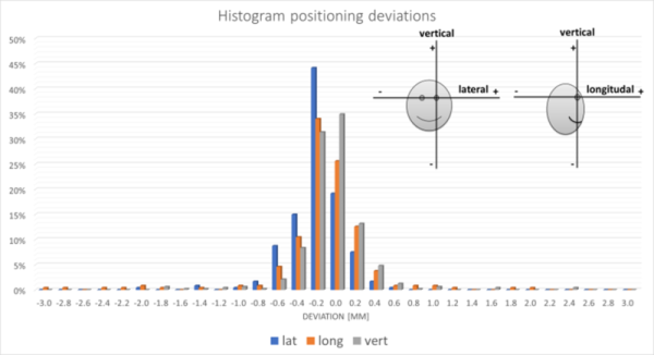
Figure 1: Histogram of found positioning deviations in OPT. Image insert
shows the patient’s reference axes.
Conclusion
The found
statistics will be used as an baseline accuracy in future work towards clipless
treatment.