Protons in posterior fossa ependymoma- a dosimetric comparison with photons
Durga Harika Kannikanti,
United Kingdom
PO-1161
Abstract
Protons in posterior fossa ependymoma- a dosimetric comparison with photons
Authors: Durga Harika Kannikanti1, Frances Charlwood1, Matthew Clarke1, Rovel Colaco1, Shermaine Pan1, Daniel Saunders1, Peter Sitch1, Nicola Thorp1, Gillian Whitfield1,2, Maria Rasool3
1The Christie NHS Foundation Trust, Proton Beam Therapy Centre, Manchester, United Kingdom; 2Division of Cancer Sciences, University of Manchester, Manchester Cancer Research Centre, Manchester Academic Health Science Centre, Manchester, United Kingdom; 3University of Manchester, School of Medicine, Manchester, United Kingdom
Show Affiliations
Hide Affiliations
Purpose or Objective
Adjuvant radiotherapy in ependymoma
improves both local control and survival outcomes. The 5 year overall survival in
paediatric ependymoma is around 85%. With such outcomes, the late effects of
treatment assume prime importance. Protons when compared with photons have the
potential to spare organs at risk (OARs), and thereby decrease late
complications. This study was designed to compare the dosimetry of proton with photon
radiotherapy plans in patients with posterior fossa ependymoma, in order to
understand the potential for reduced late effects.
Material and Methods
Data were extracted for ten
patients with posterior fossa ependymoma treated with protons who had Volumetric
Modulated Arc Therapy (VMAT) photon plans prepared in case of cyclotron
downtime. Five were single phase plans to a total dose of 59.4Gy in 33 fractions, with appropriate limitation of dose at spinal
cord level, and 5 were two phase plans, to 54Gy in 30 fractions followed by a boost of
5.4Gy in 3 fractions to the volume above the foramen magnum. Clinically relevant dosimetric variables of target and
routinely outlined OARs for data collection were selected based on our clinical
practice and with reference to the European Particle Therapy Network (EPTN)
consensus on radiation dose constraints for OARs in neuro-oncology. Mean values were compared between proton and
photon plans using a two tailed paired sample t-test. P values of less than
0.05 were considered statistically significant.
Results
Of ten patients, 7 were
females. The median age was 7 years (range 3 to 17 years). For almost all
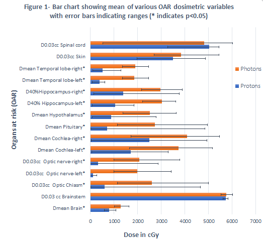
dosimetric variables, proton plans showed a statistically significant dosimetric
advantage in OAR variables, including to the optic nerves and chiasm, cochleae,
pituitary gland, hypothalamus, hippocampi, temporal lobes, brain and brainstem.
The mean D0.03cc for brainstem, skin and spinal cord were not statistically
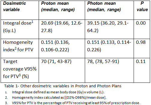
significantly different between modalities (Figure 1). The mean integral dose was
significantly lower in protons compared to photons (p=0.00) with a median proton
to photon integral dose ratio of 0.55 (range-0.41 to 0.74). The mean percentage
of the PTV receiving 95% of prescription dose and the mean
homogeneity index were not statistically significantly different between proton
and photon plans (p=0.11 and p=0.98 respectively, Table 1).


Conclusion
This study showed marked
dosimetric advantage for protons in OAR sparing and lowered integral doses,
while maintaining similar target volume coverage. This is anticipated to reduce
the risk of late effects, particularly hearing loss, endocrine dysfunction,
neurocognitive effects, and second malignancies, while maintaining the
established high rates of local control with photons.