Quantification of diaphragm motion with ultrasound during non-invasive high frequency ventilation
PO-1056
Abstract
Quantification of diaphragm motion with ultrasound during non-invasive high frequency ventilation
Authors: Kaylee van Duren1, Jeffrey Veldman1, Michael Parkes1, Zdenko van Kesteren1, Markus Stevens2, Joost van Schuppen3, Geertjan van Tienhoven1, Arjan Bel1, Irma van Dijk1
1Amsterdam UMC, Radiation Oncology, Amsterdam, The Netherlands; 2Amsterdam UMC, Anaesthesiology, Amsterdam, The Netherlands; 3Amsterdam UMC, Radiology, Amsterdam, The Netherlands
Show Affiliations
Hide Affiliations
Purpose or Objective
Breathing motion causes substantial
uncertainties in tumor position during radiotherapy. High frequency ventilation
with small tidal volumes reduces the amplitude of breathing motion. The aim of
this study was to quantify the motion of the right diaphragm dome in conscious
healthy volunteers during non-invasive mechanical ventilation without
percussion at various high frequency settings, using ultrasound.
Material and Methods
Seven volunteers were mechanically
ventilated with Positive End Expiratory Pressure (PEEP) at 60 breaths per
minute (brpm) using a Hamilton-T1 ventilator, and at 60, 150, 250 and 400 brpm
using a TwinStream jet-ventilator. During 40 s recordings, 900 consecutive sagittal
images of the diaphragm were acquired using the bk3000 ultrasound system (BK
Medical Benelux NV/SA, Mechelen, Belgium) with a convex ultrasound probe at 3.5
MHz and a temporal resolution of 23 frames per second. The same trained operator
made all measurements. Measurements were undertaken in two separate sessions. The
order of ventilation frequency settings for the TwinStream was randomized. The
images from six volunteers of both measurement sessions and at all ventilation
frequency settings were adequate and further analyzed semi-automatically in
MATLAB (n=12). Excursions were determined based on a time motion image along
the direction of movement near the anterior layer of the coronary ligament
(Fig.1). From each time motion image, the diaphragm was segmented by thresh holding
and the mean diaphragm excursion (peak – trough) for all inflations determined
and analyzed with a Wilcoxon signed-rank test (Bonferroni adjusted α=0.05/4).
Results
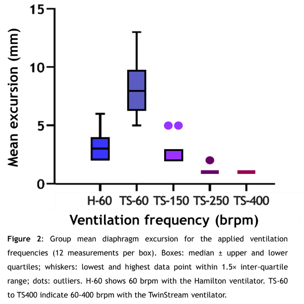
The mean diaphragm excursion (8mm)
at 60 brpm using the TwinStream ventilator showed a significantly larger median excursion
(p=0.002) than when using any other ventilation frequency setting (Fig. 2). The smallest diaphragm excursions, with a
mean of 1 mm, were found using the TwinStream ventilator at 250 and 400 brpm. At
all ventilation frequencies except using the TwinStream ventilator at 60 brpm the
mean inter-session variability was <1 mm.


Conclusion
Motion
of the right diaphragm dome during non-invasive high frequency ventilation was
successfully quantified using ultrasound. High frequency ventilation without
percussion is a promising breathing regularization strategy to reduce the
diaphragm motion, especially using the TwinStream ventilator at 250 or 400 brpm.