Evaluation of CBCT-based auto contouring for online adaptive radiotherapy of cervical cancer
Catherine Khamphan,
France
PO-1635
Abstract
Evaluation of CBCT-based auto contouring for online adaptive radiotherapy of cervical cancer
Authors: Aurelien BADEY1, Antoine Arnaud2, Ana Bigou2, Gaëtan De Rauglaudre2, Anthony Nagy1, Laure Vieillevigne3,4, Catherine Khamphan1
1Institut du Cancer Avignon-Provence, Medical Physics, Avignon, France; 2Institut du Cancer Avignon-Provence, Radiotherapy, Avignon, France; 3Institut Claudius Regaud - Institut Universitaire du Cancer de Toulouse , Medical Physics, Toulouse, France; 4Centre de Recherches en Cancérologie de Toulouse, UMR1037 INSERM - Université Toulouse 3 – ERL5294 CNRS, Toulouse, France
Show Affiliations
Hide Affiliations
Purpose or Objective
Daily online Adaptative RadioTherapy (oART) sessions requires new organs at risk (OAR) and target delineation. oART Cone-Beam CT (CBCT)-based available on ETHOS Therapy system uses AI segmentation and deformable image registration (DIR) for OAR and target volumes in pelvic region. This work evaluates the contouring accuracy obtained and its impact on dose metrics.
Material and Methods
Five patients already treated on Halcyon system for cervix cancer (45Gy/25 fr.) in our clinic were randomly selected, 10 oART sessions per patient were simulated on different CBCT using an ETHOS emulator. Each CBCT was manually delineated offline by an expert, determining the reference contours (ref) which were subsequently compared to contours generated from ETHOS uncorrected (ETHOSno,corr) and with manual correction (ETHOScorr).
The study included contours evaluation with comparison metrics (DICE index, Mean Distance to Conformity (MDC)) and dosimetric evaluation using corrected and uncorrected contours from oART sessions. A complementary qualitative contours evaluation was carried out with a likert scale (0: accepted, 1: accepted with minor modifications, 2: accepted with major modifications, 3: rejected).
Results
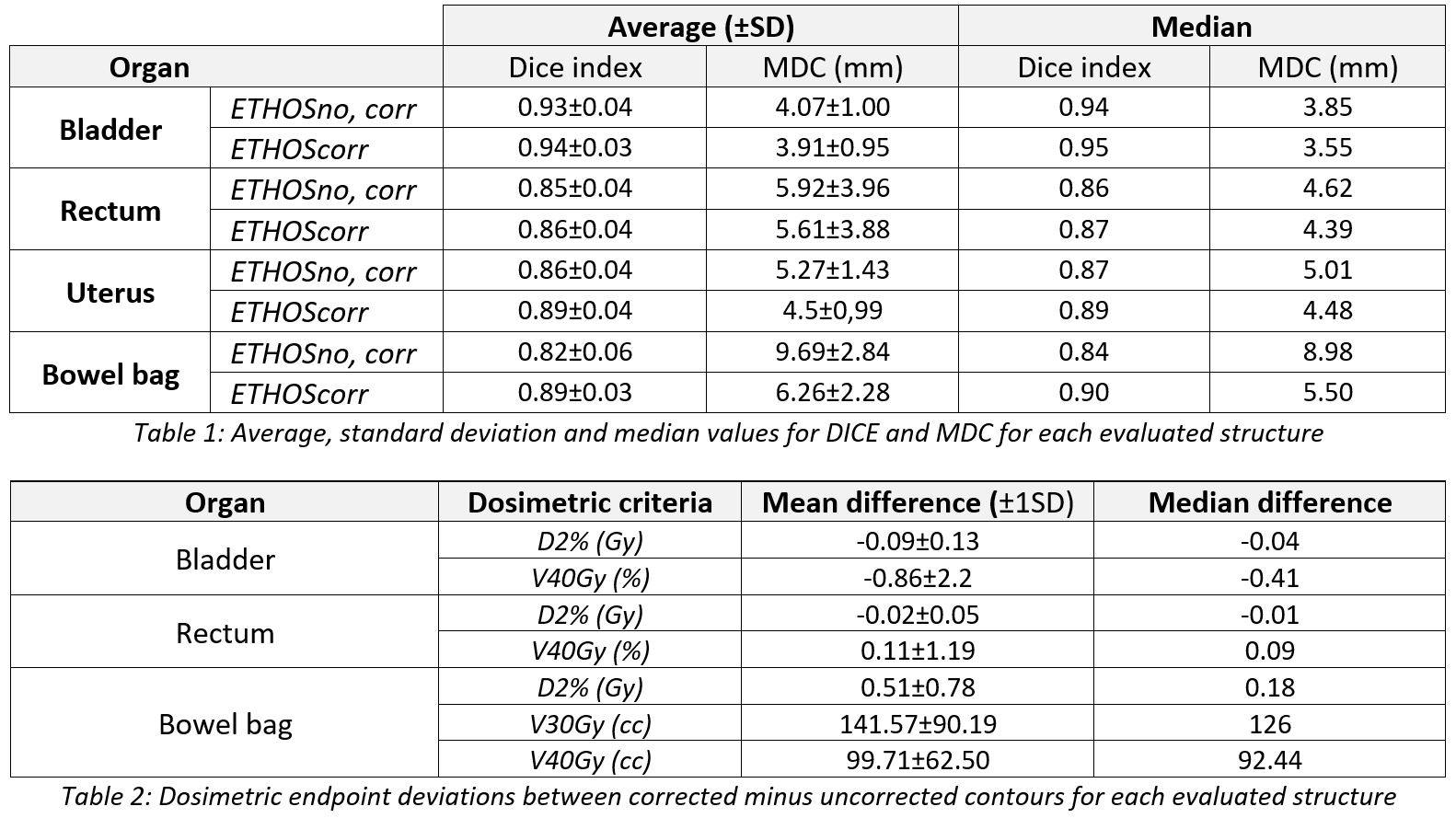
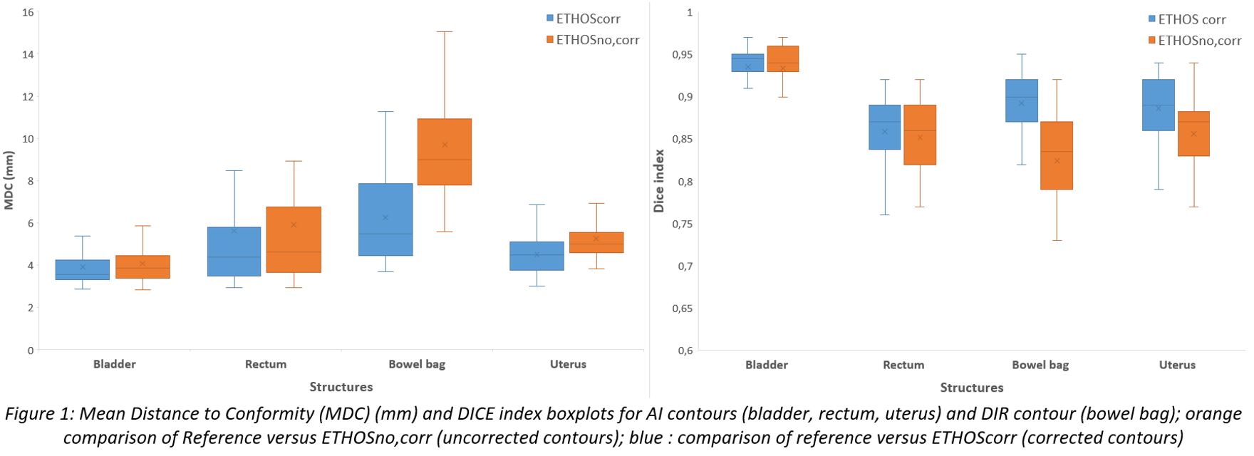
Bladder delineation shows better results, while uterus and rectum have acceptable values despite a greater dispersion. The most important differences are observed for bowel bag which is the only structure obtained by DIR (Table 1). Greater dispersion is observed for patient 1 on bladder and bowel bag due to CBCT image quality and presence of gas (Figure 1). The dosimetric impact of using corrected or uncorrected contours is highlighted in Table 2 and is the most remarkable for bowel bag.


Dosimetric criteria are all respected for bladder and rectum whether the corrected or uncorrected contours are used. For bowel bag, however, it appears necessary to adjust the contours manually (online or offline). The qualitative evaluation shows that none of the contours provided by ETHOS were rejected, the majority were adjusted.
Conclusion
The evaluation shows excellent results for a majority of AI contours with CBCT images. The impact of manual correction on ETHOS contours is significant in terms of improvement of metrics, but the dosimetric impact is only significant for bowel bag, implying the need to recontour this structure during (or after) the oART CBCT-based session. Additional work is underway to optimize CBCT image quality.