Performance characterization of the Blue Physics scintillator in the 1.5T MR-linac
Stijn Oolbekkink,
The Netherlands
PD-0588
Abstract
Performance characterization of the Blue Physics scintillator in the 1.5T MR-linac
Authors: Stijn Oolbekkink1, Bram van Asselen1, Simon Woodings1, Jochem Wolthaus1, Wilfred de Vries1, Arian van Appeldoorn1, Marcos Feijoo2, Madelon van den Dobbelsteen1, Bas Raaymakers1
1UMC Utrecht, Radiotherapy, Utrecht, The Netherlands; 2Blue Physics, Inc., Lutz, USA
Show Affiliations
Hide Affiliations
Purpose or Objective
On the MR-linac, the treatment is adapted based on the daily anatomy. Dosimetric plan QA is usually based on integral dose verification. However, to verify real-time adaptive treatments, they should be measured with a high temporal resolution. Furthermore, the detectors should not be affected by, or distort, MR image acquisitions used to measure full adaptive workflows, including cine imaging for motion monitoring. As plastic scintillating dosimeters (PSD) potentially have these properties, we have tested the performance of the Blue Physics (Lutz, FL) PSD system for use in the Elekta 1.5T MR-linac (Stockholm, SE).
Material and Methods
Besides the dose signal obtained from the scintillator in the PSD, Cherenkov radiation is generated by fast electrons inside the plastic fiber. To detect the amount of Cherenkov signal, a second adjacent fiber, without scintillator, is used. Several calibration procedures were tested in both a conventional linac and MR-linac.
Performance of the PSD was tested in terms of detector stability, dose linearity, dose rate stability, axial rotation, PDD and output factors. Measurements were performed in a PTW BEAMSCAN MR (Freiburg, DE) water phantom with a PTW microDiamond as reference detector. Furthermore influence of the orientation of the detector relative to the magnetic field was determined using a PTW RW3 slab phantom.
The application of patient plan verification was tested by irradiating an IMRT plan on the RTsafe Prime head phantom (Athens, GR). A comparison was made to the same plan measured with a PTW Semiflex 0.125cc detector.
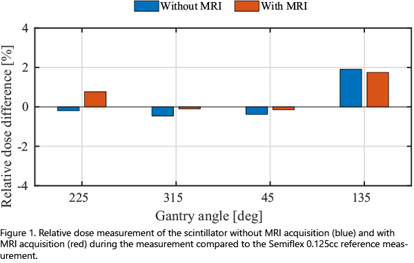
MRI acquisition dependence was tested with the Modus QUASAR MRI 4D motion phantom (London, ON, CA). Experiments were conducted with the PSD and Semiflex for gantry angles 225°, 315°, 45° and 135° with and without MRI acquisitions.
Results
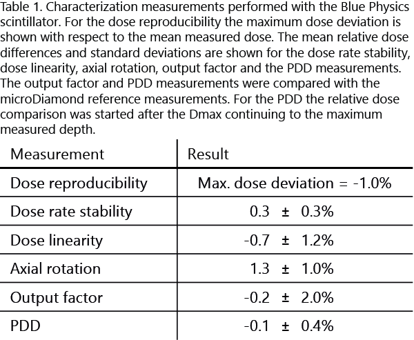
The various calibration methods showed a larger variation in calibration factors on the MR-linac relative to a conventional linac. The results of basic characterization tests are shown in Table 1. PDD showed good correspondence with the microDiamond detector. The response of the detector was influenced by the orientation of the detector relative to the magnetic field up to 10.7%.

Plan QA showed similar results for the PSD and Semiflex measurement with a dose difference of 0.6%.
The dose differences with and without MRI acquisitions are plotted in Figure 1. Relative dose differences and standard deviations were 0.6±0.9% and 0.2±1.1%, respectively, for with and without MRI acquisition relative to the Semiflex measurement.

Conclusion
Basic performance of the Blue Physics PSD has been tested and shown to be suitable for relative dosimetry in the MR-linac. The calibration methods showed variation and need to be further investigated. Furthermore the detector is dependent on the orientation relative to the magnetic field. No significant difference in dose measurements with and without MRI acquisition were observed. Further investigation is needed to qualify as an absolute dosimeter for the MR-linac.