An evaluation of the suitability of decorated immobilisation in MRI and Proton Beam Therapy
Jennifer O'Brien,
United Kingdom
PO-2256
Abstract
An evaluation of the suitability of decorated immobilisation in MRI and Proton Beam Therapy
Authors: Jennifer O'Brien1, Callum Gillies2, Claire Hardy1
1University College London Hospitals, Radiotherapy/Proton Beam Therapy, London, United Kingdom; 2University College London Hospitals, Proton Beam Therapy, London, United Kingdom
Show Affiliations
Hide Affiliations
Purpose or Objective
The immobilisation shells for paediatric patients having radiotherapy at UCLH are decorated by an artist to make them more ‘child-friendly’. In photons at UCLH, painting these shells is standard practice, using liquid acrylic paints.
In the PBT department, verification MRI scans are done weekly for patients with diagnoses whereby anatomical change is expected. To establish a standard practice across protons and photons, the original paints and an alternative were tested to ensure appropriateness.
An alternative paint was sourced, (paint pens from POSCA™). To ensure these were appropriate for PBT and MRI, they were analysed by radiographers and PBT physics, ensuring the outcome was still suitable for purpose (evaluated by PBT play specialists).
The original standard paints were evaluated versus POSCA™ paint pens.
Two evaluations were done to establish:
• The effect of the paint used on MR imaging
• That the Water Equivalent Thickness (WET) of decorated Q-Fix™ BoS immobilisation shells is modelled sufficiently by PBT planning systems
Material and Methods
The first evaluation determined if the paints had any the effect on MRI image quality. A homogenous phantom was scanned on the Phillips Ingenia 1.5T MRI with an immobilisation mask decorated with standard liquid acrylic paints fixed around it, followed by one decorated with POSCA™ pens. A T2 sequence from the departmental Head and Neck protocol was used and images then evaluated by radiographer and physics staff.
The second evaluation aimed to ensure that the WET modelled by Eclipse matched measured values on the treatment unit using an MLIC (Multi-layer Ionisation Chamber). The decorated shell was scanned using the Head and Neck protocol on the PBT CT scanner and imported to Eclipse. The WET was measured with the tools available in Eclipse in a variety of places where physical measurements could be taken on set using the MLIC. A 160MeV beam was shot into the MLIC as a reference. The distal 80% fall off of the Bragg peak was measured with and without theses mask sections in the beam path. The difference in the measured D80 then represented the measured WET of that mask region.
Results
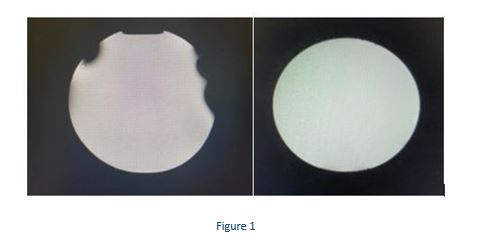
The MRI image on the left of Figure 1 shows significant artefact given by the standard liquid acrylic paints, deeming them inappropriate for use in PBT due to their lack of compatibility with MRI. Opposingly, the right of Figure 1 shows the phantom with a mask painted with POSCA™ paints around it and shows no artefact from this. If these paint pens would have no impact on dosimetry, these looked to be the better solution.

In the second evaluation, both sections of the painted mask had D80 values of 17.47cm which means a measured WET of 0.14cm, lying within the values measured in Eclipse.

Conclusion
The POSCA™ pens used to decorate immobilisation shells do not cause issues with the quality of MRI scans and can be appropriately modelled by the planning system for accurate dosimetry. The service is recommended to only use POSCA™ pens to decorate immobilisation.