Quantification of pelvic rotations of gynecological cancer patients positioned with surface imaging
Mimmi-Caroline Bolin,
Sweden
PO-1923
Abstract
Quantification of pelvic rotations of gynecological cancer patients positioned with surface imaging
Authors: Mimmi-Caroline Bolin1, Marianne Falk1, Mattias Hedman2, Giovanna Gagliardi1, Eva Onjukka1
1Karolinska University Hospital, Medical Radiation Physics and Nuclear Medicine, Stockholm, Sweden; 2Karolinska University Hospital, Radiation Oncology, Stockholm, Sweden
Show Affiliations
Hide Affiliations
Purpose or Objective
The aim of this study was to determine if rotational uncertainties for gynecological cancer patients can be reduced by surface imaging (SI), as compared to aligning three markers on the patient body with in-room lasers (marker-laser). As rotational deviations from the planned position can have severe impact on the set-up accuracy for patients with the para-aortic lymph nodes (PALN) included in the CTV, the quantification of positioning accuracy is of high importance.
Material and Methods
Fifty gynecological patients treated with external radiotherapy were retrospectively included in this study, 25 patients positioned with marker-laser and 25 patients positioned with SI. The values of rotational (pitch and roll) deviations of the patient positions between treatment planning CT (TPCT) and online CBCT were collected for both subcohorts, and all treatment fractions, after performing an automatic registration between the two image sets. The statistical analysis of the difference between the two set-up methods was done with the Mann-Whitney U-test. Yaw rotation and translations were not included in the quantification of positioning accuracy as those parameters were corrected for in the online registration between TPCT and CBCT. Each patient that required re-imaging was recorded, as an indication of large residual set-up errors after initial set-up. For all patients, the extent of the CTV was measured in the cranio-caudal (CC)-, anterior-posterior (AP)- and lateral-medial (LM) directions, from the isocenter to the most distant point on the contour, to allow quantification of the potential impact of errors, in pitch and roll, on target misalignment.
Results
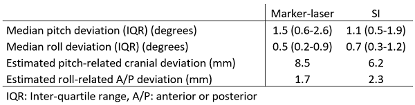
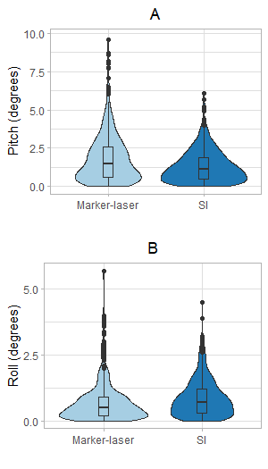
The absolute pitch deviation was greater for the marker-laser method than for the SI method (p<0.01), while the absolute roll deviation was smaller for the marker-laser method than for the SI-based method (p<0.01). Median and inter-quartile range values are presented in the table, and the figure shows the absolute distributions of the rotational deviations. However, since rotational deviations result in increasing misalignment further from the isocenter, pitch deviations have a greater impact on the accuracy of the set-up. For a patient with PALN CTV (CC length of 18.8 cm and AP depth of 10.9 cm) the pitch-related vertical deviation at the cranial end of the target is much greater than the corresponding roll-related longitudinal deviation at the outer A/P location, estimated based on the upper end of the IQR for rotational deviations (see table).
Fewer patients (10/25) needed to be re-imaged when using SI, compared with marker-laser (17/25) as positioning method.
Conclusion
By introducing SI as a set-up method for gynecological cancer patients, higher positioning accuracy can be achieved compared to the marker-laser set-up method.