Development and test of a patient immobilization and rotation device for imaging and radiotherapy
PO-1913
Abstract
Development and test of a patient immobilization and rotation device for imaging and radiotherapy
Authors: Gernot Echner1,2, Fabian Dinkel1,2, Cedric Beyer3, Oliver Jäkel1,4,5
1German Cancer Research Center (DKFZ), Medical Physics in Radiation Oncology, Heidelberg, Germany; 2National Center for Radiation Research in Oncology (NCRO), Heidelberg Institute for Radiation Oncology (HIRO), Heidelberg, Germany; 3University Hospital Heidelberg, Radiation Oncology, Heidelberg, Germany; 4National Center fpr Radiation Research in Oncology (NCRO), Heidelberg Institute for Radiation Oncology (HIRO), Heidelberg, Germany; 5University Hospital Heidelberg, Heidelberg Ion Beam Therapy Center (HIT), Heidelberg, Germany
Show Affiliations
Hide Affiliations
Purpose or Objective
Radiotherapy is one of the most prescribed tumor treatments, using high doses of ionizing irradiation to achieve tumor control and preserve healthy tissue from damage. As there may be significant differences between patient’s morphology at the treatment day compared to the day of the planning we designed a new principle to acquire daily MR images for any final treatment position to enable irradiation without a gantry. Therefore, a patient fixation and rotation system was developed and tested for CT and MR scanners, as well as on the treatment table of a light ion beam treatment facility.
Material and Methods
The rotation system was designed with Inventor 2018 (Autodesk, San Rafael, USA) and some bearing parts were 3D-printed with the Objet 500 Connex3^TM printer (Stratasys, Israel) using VeroCyan^TM. For additional plastic and wooden parts, manufacturing was realized at different workshops and worm-gear-boxes were bought (igus® GmbH, Köln). All parts were assembled in-house at the DKFZ and tested during a clinical study with 19 subjects at the university clinic Heidelberg
Results
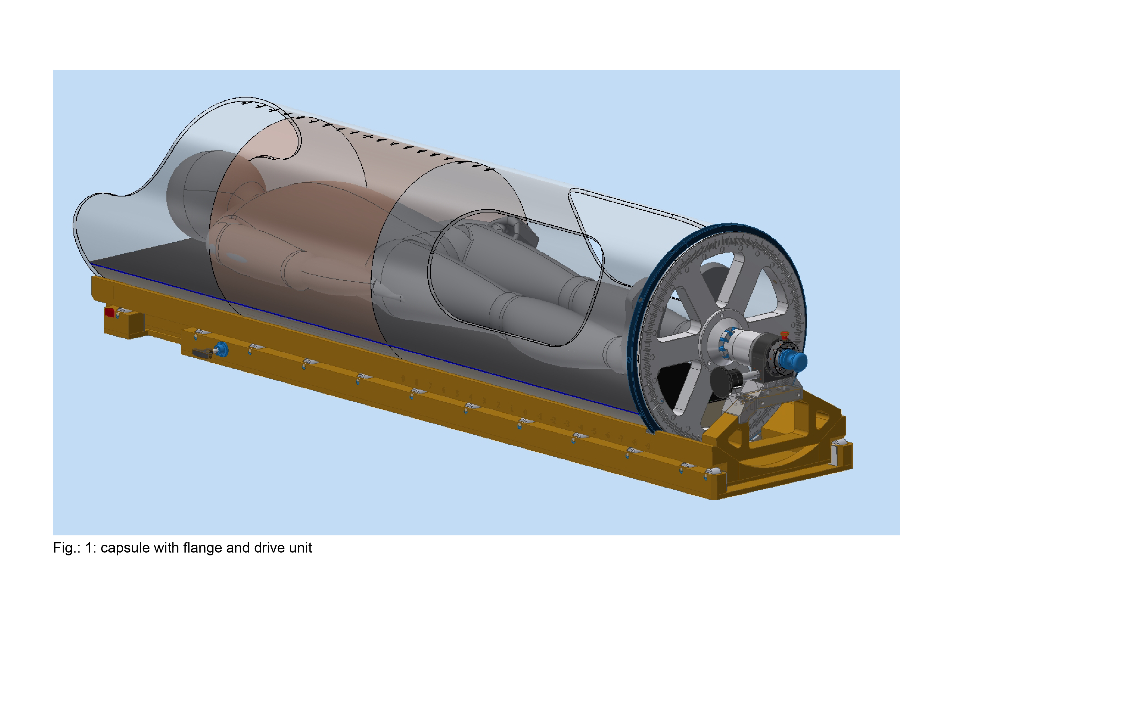
The most challenging requirement for the rotation system was to abstain from metal material as the device is used inside an MR scanner. Therefore, mostly plastics and wooden materials were used. Additional requirements were the use of homogeneous material in the treatment area of the body without any air gap in the beam area. This is achieved by using an acrylic glass tube with an outer diameter of 550mm and a wall thickness of 10mm to fit into the MRI. This tube is pivot-mounted in a baseplate which fits into the MRI. A bottom side mounted flange can be manually driven by two worm gears. One of the gears is used to rotate the subject in standard speed, the second one can be connected for fine adjustment and is also used as a break to fix this position. The subject is fixed in-between 2 vacuum mattresses and moved axially inside the tube (Fig.:1). After that the arrangement is carried into the MRI room and then moved into the scanner to a pre-defined position. Positioned in the MRI, the subject can be rotated with the capsule to the desired angle. So far, 19 subjects were tested and did not interrupt the test procedure of about 50 minutes.
Conclusion
A patient immobilization and rotation system was developed and tested to enable MR and CT imaging directly before irradiation to adapt the treatment plan according to the daily tumor and OAR position and shape. Additionally, this setup enables radiation treatment from different angles even at a fixed horizontal beam-line, as the capsule can be rotated axially on the treatment table. Thereby, the need for an expensive gantry for irradiation becomes obsolete. This may allow for much cheaper installation of treatment facilities with high quality image guidance. After receiving the CE labelling we will finally test the system with patients in the near future also in a CT environment. In the long term, the system may also be used for a low-cost MR-Linac solution.