Vienna, Austria

ESTRO 2023

Session Item

Inter-fraction motion management and offline adaptive radiotherapy
7006
Poster (Digital)
Physics
Predicting organ motion in cervix radiotherapy: unlocking the secrets of the planning scan
Lei Wang, United Kingdom
PO-1911

Abstract

Predicting organ motion in cervix radiotherapy: unlocking the secrets of the planning scan
Authors:

Lei Wang1, Dualta McQuaid1, Sarah Mason2, Matthew Blackledge2, Helen McNair1, Emma Harris2, Susan Lalondrelle3

1Royal Marsden Hospital NHS Trust, Radiotherapy department, Sutton, United Kingdom; 2Institute of Cancer Research, Department of Radiotherapy and Imaging, Sutton, United Kingdom; 3Royal Marsden Hospital NHS Trust and Institute of Cancer Research, Department of Radiotherapy and Imaging, Sutton, United Kingdom

Show Affiliations
Purpose or Objective

Motion of the low-risk clinical target volume (CTV-LR) during cervix cancer radiotherapy can be extensive and complex. Motion of the uterocervix has been documented to relate to bladder and rectal size, but predictive models based on these alone have been imperfect. We aim to create a model that describes and predicts interfractional motion on an individualised basis, using anatomical, pathological and demographical features.

Material and Methods

160 images from 20 cervix cancer patients who received radical radiotherapy were evaluated. For each patient, the full-bladder CT, empty-bladder CT, planning MRI and 5 cone-beam CTs chosen at random were contoured (CTV-LR, bladder, rectum) following the EMBRACE-II protocol. In addition, 23 features of the planning CTs and patient demographics were measured, including various measures of organ shape, and contents, and analysed for their influence on subsequent motion of points U (tip of uterine fundus), C (posterior cervix) and center of mass of CTV-LR (CoM) assessed using linear regression and Wilcoxon signed-rank test.

Results

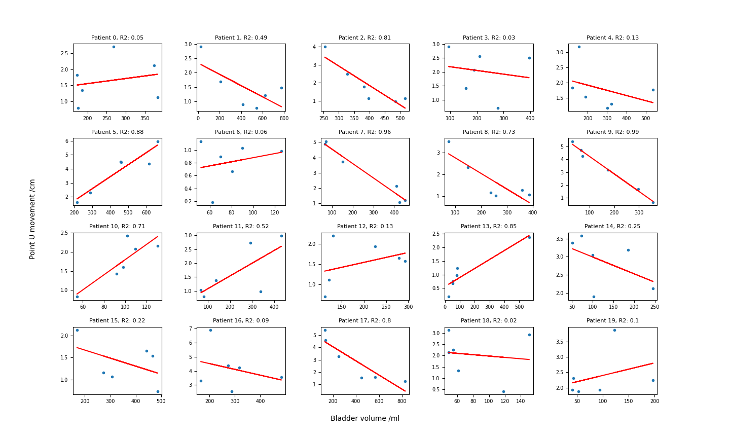
Variation was observed in bladder volumes (mean 239 ml, range 25-829), rectal diameters (mean 2.6 cm, range 1.1-5.7) and euclidean distance moved by U (mean 2.2, range 0.2-6.9), C (mean 1.3 cm, range 0.2-3.3), and CoM (mean 1.0 cm, range 0.1-3.1). For the overall cohort, bladder volume and rectal diameter did not correlate with distance moved by U, C or CoM (R2 all < 0.1). However, some individuals had stronger correlations; for U motion against bladder volume, 8 patients had R2 > 0.7 (Figure 1).

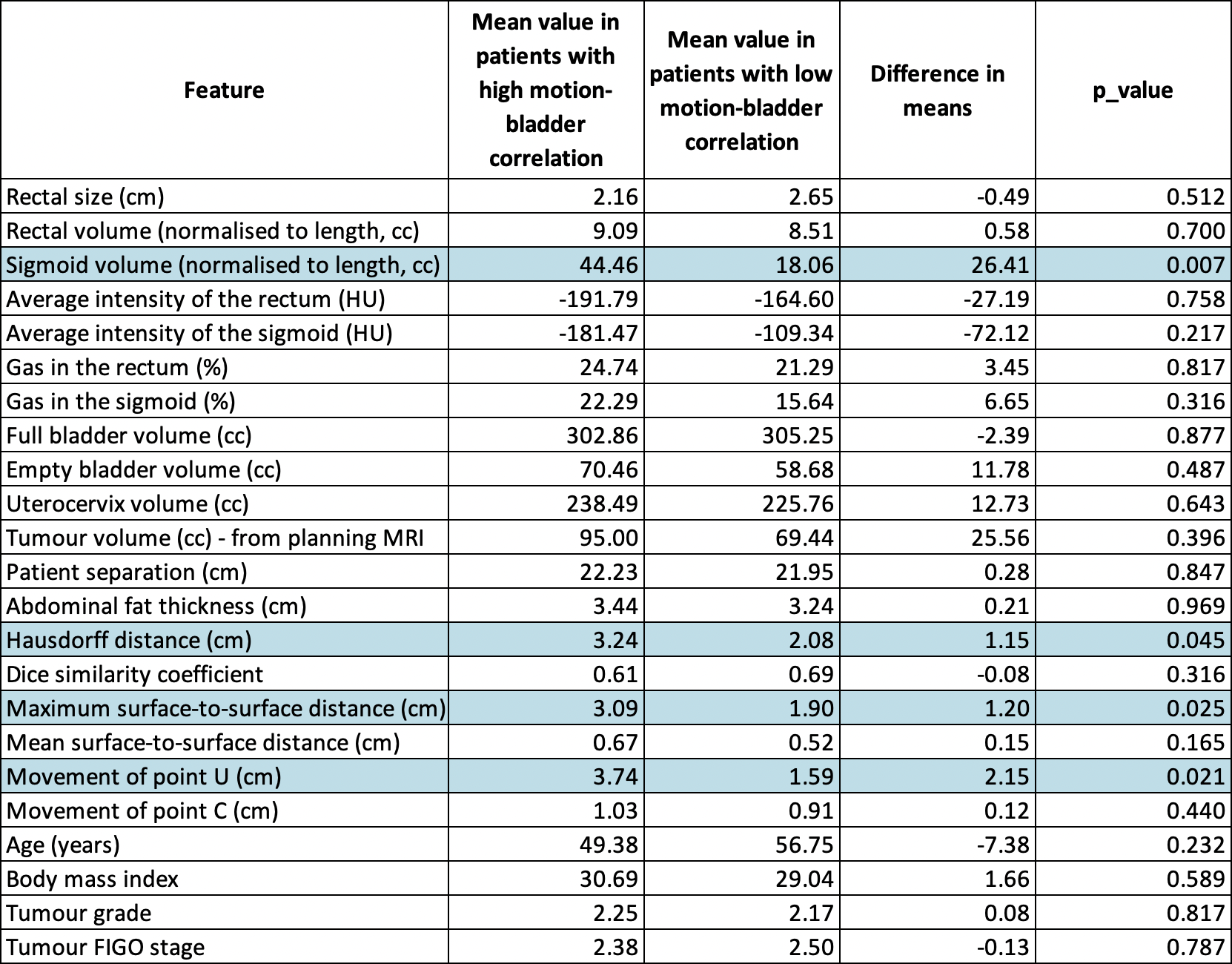
Patients with a stronger bladder-motion correlation had larger sigmoid volume on CT (p = 0.006) and more motion of CTVLR between full and empty-bladder CTs (more motion of the uterine fundus tip, p = 0.020; larger maximum surface-to-surface distance, p = 0.025; larger Hausdorff distance, p = 0.044). Details in Table 1.

The slope of the relationship could be predicted pre-treatment based on sigmoid volume at planning, difference in bladder volume between full and empty CTs, tumour volume on MRI, and change in the superior extent of the CTVLR contour between full and empty planning CTs (linear regression, R2 0.77). Rectal size generally had weaker per-patient correlations with U, C and CoM. When bladder and rectal size were combined as explanatory variables, U motion could be predicted with confidence (R2 > 0.7) in 10 patients, C motion in 7 patients, and CoM motion in 8 patients.

Conclusion

Interfraction target motion during cervix cancer radiotherapy is dependent on bladder volume and rectal diameter, but the strength of correlation and slopes vary by patient. We have demonstrated that several features that influence this relationship can be extracted from the planning CT. Further efforts to develop a predictive motion model could provide valuable prior knowledge for adaptive radiotherapy strategies including automatic plan of the day selection and online replanning.