An evaluation of a novel CBCT system: image quality, extended FoV and metal artefact reduction
Gabriel Paiva Fonseca,
The Netherlands
PO-1668
Abstract
An evaluation of a novel CBCT system: image quality, extended FoV and metal artefact reduction
Authors: Gabriel Paiva Fonseca1, Didier Lustermans1, Marta Bogowicz1, Vicki Taasti1, Femke Vaassen1, Richard Canters1, Koen Theeuwen1, Wouter van Elmpt1, Frank Verhaegen1
1Maastricht University Medical Centre+, Department of Radiation Oncology (MAASTRO), GROW – School for Oncology and Reproduction, Maastricht, The Netherlands
Show Affiliations
Hide Affiliations
Purpose or Objective
A novel cone-beam CT (CBCT) imager including advanced reconstruction capabilities was developed for optimized radiotherapy workflow. This study evaluates this CBCT (HyperSightTM integrated on a Halcyon v4.0 (pre-market release), Varian Medical Systems) for image quality of fast CBCTs (acquisition time of 5.9 s) and novel features such as extended FoV (eFoV) up to 70 cm and two reconstruction techniques (iCBCT Acuros and iCBCT MAR for metal artefacts reduction) introduced in this version.
Material and Methods
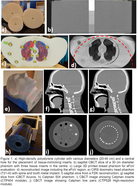
CBCTs were acquired using multiple commercial phantoms (Figure 1) and reconstructed using FDK and iterative reconstruction (iCBCT Acuros and iCBCT MAR). HU accuracy was evaluated using tissue-mimicking inserts (Gammex, Sun Nuclear) in multiple positions within anthropomorphic phantoms and cylindrical phantoms with diameters from 20 up to 40 cm. A large 3D-printed breast phantom designed for eFoV was used to evaluate geometrical and HU accuracy outside (up to 70 cm) of the standard FoV (54 cm; sFoV). In addition, image resolution and contrast were also evaluated using Catphan and MAR was evaluated using a CIRS head phantom with tooth and metal spine implants. Results were compared with fan-beam CT images (Siemens SOMATOM Drive).
Results
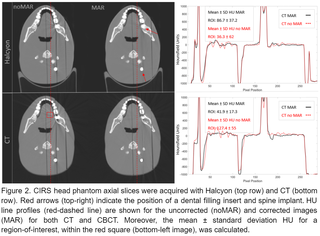
The spatial resolution measured with the Catphan phantom results showed spatial resolution well within 0.45-0.60 lp/mm and overall uniformity above 98.3% for all protocols. Iterative reconstruction resulted in fewer image artefacts and noise (see Figure 1 f-g). HU variation due to the phantom was within 30 HU for most of the materials and 80 HU for cortical bone when comparing a large diameter phantom (40 cm diameter) against the 20 cm one. The effect of the eFoV on the HU is more pronounced, due to the necessary use of truncated data, with better agreement for lung and soft tissues. The geometrical accuracy of the eFoV reconstruction depends on the volume within the eFoV being of the order of a few millimetres for most of the regions. The effect of the metal artefact reduction is shown in Figure 2 where iCBCT MAR shows reduced artefacts when compared to the image without correction and image quality compatible with a CT acquisition.
Conclusion
This study presents an extensive validation of a pre-market release of the novel CBCT imager. The variety in size, materials and other phantom features allowed qualitative and quantitative evaluations of multiple imaging features. The novel CBCT panel has a large sFoV and additional eFOV capability. Although more pronounced variations were observed for the HU values of tissue-mimicking inserts in the eFoV, geometrical accuracy is comparable to standard planning CT eFoV reconstruction.