Histopathological examination of proton radiation-induced late effects in mice legs
Cathrine Bang Overgaard,
Denmark
PO-2205
Abstract
Histopathological examination of proton radiation-induced late effects in mice legs
Authors: Cathrine Bang Overgaard1, Danny Mortensen2, Jens Randel Nyengaard2,3, Trine Tramm2, Brita Singers Sørensen1,4
1Aarhus University Hospital, Experimental Clinical Oncology, Aarhus, Denmark; 2Aarhus University Hospital, Pathology, Aarhus, Denmark; 3Aarhus University, Core Center for Molecular Morphology, Section for Stereology and Microscopy, Clinical Medicine, Aarhus, Denmark; 4Aarhus University Hospital, Danish Centre for Particle Therapy, Aarhus, Denmark
Show Affiliations
Hide Affiliations
Purpose or Objective
Radiation-induced fibrosis is a common chronic and progressive side effect post radiotherapy. It is important to understand the radiobiological rationale for chronic effects of proton radiation to improve the quality of life of multiple patients. The challenge is to compare the observable changes with the actual pathological tissue changes post radiation. The purpose of this exploratory study was to test a procedure for histochemical and stereological examination of proton-irradiated mice legs that received different doses and compare this to a leg contraction assay.
Material and Methods
The right hindlimb of 4 mice were irradiated with a single high PBS proton dose of 27 Gy, 31 Gy, 33 Gy or 45 Gy. They exhibited different degrees of late effect severity in a previously published leg contraction assay (score 2: low, score 3: medium, score 4: severe). The treated right hindlimb and the control left leg were harvested post one year of functional late effect follow up. Three cross sections of the legs were cut, namely the upper leg (above ancle joint), the ancle joint and the foot. The tissue sections were stained with Hematoxylin-Eosin or Masson’s Trichrome to evaluate their total area, area fraction of connective- and adipose tissue, epidermal thickness, and total skin adnexa (hair follicles and glands) profile count. Area fraction was evaluated using stereological point sampling. Epidermal thickness was measured from the basal layer up to and including the granular layer with orthogonal intercept length. Total tissue section area was estimated by 2-dimensional nucleator.
Results
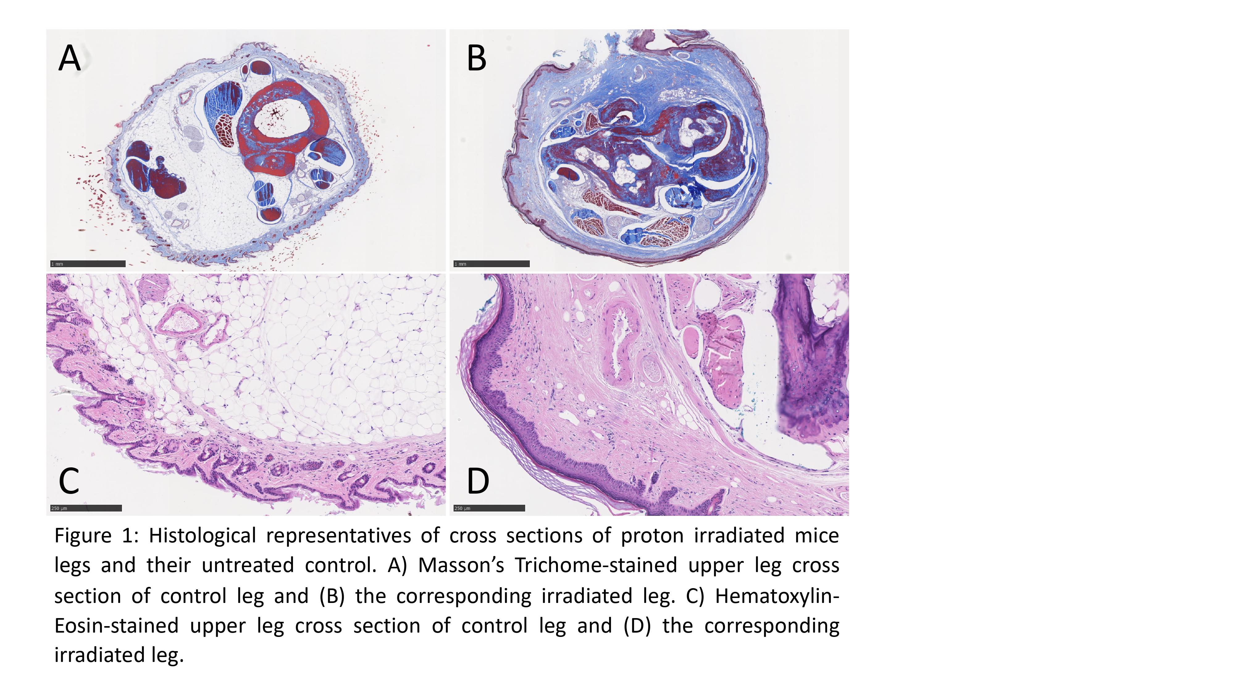
Loss of total tissue area, reduced adipose tissue fraction, increased epidermal thickness and almost complete skin adnexa ablation were all characteristics of the irradiated legs. Histological representatives are presented in Figure 1. Generally, the fraction of connective tissue was primarily increased in the upper leg sections, whereas loss of adipose tissue and total tissue area was observed in upper leg and the joint as well. The most severe changes were seen in the epidermal thickness and almost total ablation of the skin adnexa across all three sections. Mice with the most severe degree (score 4) in the functional joint flexibility assay also possessed most structural alterations.
Conclusion
Fundamental structural changes occur in mice legs post proton therapy. Clinical findings of leg atrophy (loss of total area and hair) and decreased flexibility could be visualized and quantified using stereology assisted histopathology. Evaluation in a larger study with stratification according to radiation dose is needed to investigate if a dose-response relationship can be supported histologically. The tested procedure for histological examination was found to be technically feasible and is expected to produce a robust pathological comparison to a functional leg contraction assay for radiation-induced late effects assessment in vivo.