Evaluation of MOSFET response in phantom for in vivo dosimetry in high-rate brachy breast treatments
PO-2183
Abstract
Evaluation of MOSFET response in phantom for in vivo dosimetry in high-rate brachy breast treatments
Authors: Marina Gutierrez Ruiz1, Jorge Alonso1, Guillermo Camacho1, Ana Reguilon1, Javier Albendea2, Frandeina Pinto1, Mara Garcia1, Ana Rivero1, Corro Uriel1, Veronica Cañon1, Maria Ferri1, Javier Anchuelo1, Rodrigo Astudillo1, Diego Bruzos1, Fernando Gomez1, Andres Vazquez1, Ignacio Raba1, M Teresa Pacheco1, Pedro J Prada1, Samuel Arrebola1, Rosa Fabregat1
1Hospital Universitario Marques de Valdecilla, Radiation Oncology, Santander, Spain; 2Hospital Universitario Marques de Valdecilla, Oncology Radiation, Santander, Spain
Show Affiliations
Hide Affiliations
Purpose or Objective
The proposed recommendations by the ICRP and the IAEA and the growing tendency to hypofractioned dose schedules in high-dose rate brachytherapy (HDR-BT) treatments lead us to require in vivo dosimetry (IVD) as an independent and patient-specific quality assurance method to evaluate the delivered dose.
A template was manufactured to perform IVD in HDR-BT breast cancer treatments with microMOSFET detectors, in a previous study [1]. Calibration and correction factors were proposed to minimize their angular, temperature, and source-detector distance dependence. The aim of this work is to evaluate the methodology proposed to perform IVD in this type of treatments in a phantom.
Material and Methods
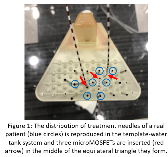
Three real HDR-BT breast treatments are reproduced on the 3D-printed template, fixed inside a water tank. The treatment needles are connected to the Elekta Flexitron afterloader with a 192-Ir source. Following the calibration geometry, three additional needles are inserted in the middle of an equilateral triangle of the treatment one for the microMOSFET detectors TN-502RDM of Best Medical Canada (figure 1).
A CT scan is performed on each distribution, the needles are reconstructed and a treatment plan similar to that of real patients in the Treatment Planning System (TPS) Oncentra Brachy is calculated. The treatment plan is delivered and the measured dose by microMOSFETs, which is the product of its calibration factor in cGy/mV and its read signal or voltage, is recorded.
The difference between the measured dose by the microMOSFETs and the calculated dose in the TPS is evaluated.
Results
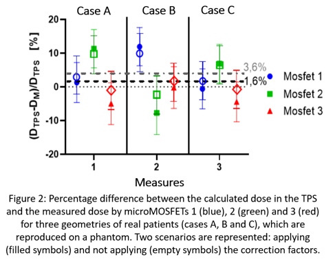
Figure 2 shows the percentage difference between measured dose by three microMOSFETs and calculated dose in the TPS for three real HDR-BT breast treatments (cases A, B and C).
The average percentage difference between measured and calculated dose with and without applying correction factors is 1.5±5.8% and 3.6±5.6%, respectively. The response of the microMOSFETs underestimates the calculated dose in the TPS and the correction factors improve the results in a 2%.
Conclusion
The proposed method to perform IVD in HDR-BT breast cancer treatments with microMOSFETs is consistent with the results of bibliography [1, 2]. Therefore, it can be carried out in clinical practice in order to monitor the delivered dose. In future studies, the IVD methodology should be evaluated in real patients in order to establish error detection thresholds to distinguish between dosimetric deviations and false alarms.
REFERENCES
[1] RUIZ-ARREBOLA, S. et al. Characterization of microMOSFET detectors for in vivo dosimetry in high-dose-rate brachytherapy with 192-Ir. Medical Physics, 2020, 47 (5), 2242-2253.
[2] RAMASESHAN, R. et al. Performance characteristics of a microMOSFET as an in vivo dosimeter in radiation therapy. Phys. Med. Biol., 2004, 49, 4031-4048 p.