Commissioning of new rectal applicator using electronic brachytherapy source
PO-2166
Abstract
Commissioning of new rectal applicator using electronic brachytherapy source
Authors: Nada Tomic1, LiHeng Liang1, Azin Esmaelbeigi2, Jonathan Kalinowski2, Shirin Enger2, Te Vuong1, Slobodan Devic3
1Jewish General Hospital, McGill University, Radiation Oncology, Montreal, Canada; 2McGill University, Medical Physics Unit, Montreal, Canada; 3Jewish GeneralHospital, McGill University, Radiation Oncology, Montreal, Canada
Show Affiliations
Hide Affiliations
Purpose or Objective
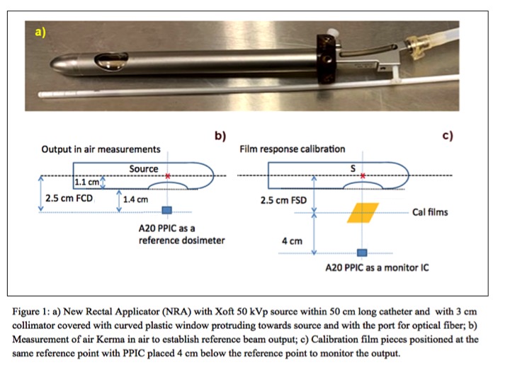
The Xoft Axxent electronic brachytherapy source with 50 kVp miniature X-ray tube is currently used to treat patients with rectal carcinoma by mimicking Papillon approach with radiation emitted through the rectoscope-like applicator (“end-on” geometry), which is uncomfortable for patients and might result in patient movement and in treatment miss. We developed a Xoft New Rectal Applicator (NRA) with side-on collimator (Fig. 1a), which allows for supine treatment position and live tumor monitoring with optical cable. We present results of measured NRA dose rate and dose distribution uniformity.
Material and Methods
We determined beam quality of the NRA with Xoft 50 kVp source by measuring HVL with parallel-plate ion chamber (PPIC) under narrow beam geometry.
We first performed relative dose measurement using EBT3 model GAFCHROMICTM film for NRA with single source position. Then, we simulated dose distribution for multiple source positions to find dwell positions and times with best achievable dose uniformity across the NRA collimator. We used air Kerma in air based reference EBT3 model film dosimetry system to estimate 2D dose distribution and the output. We performed the reference air Kerma in air measurements (Fig.1b), using calibrated Exradin A20 PPIC at reference point (AAPM TG-61). At the same point, film pieces were irradiated (Fig.1c) to create calibration curve, air Kerma in air as a function of film response ((PV0/PV) -1), which (using green color channel) was found to be linear.
In-phantom treatment plan measurement setup schematic is presented in Fig.2a. The dose distribution was measured using wrapped film pieces around NRA (Fig.2b). The in-phantom NRA output measurements were performed using PPIC (Fig.2c). Dose to water, for both measurements, was determined (AAPM TG-61) by multiplying measured air kerma in phantom with (μen/ρ)waterair.
Results
For the described NRA system, the measured HVL is 0.5 mm Al. For the created radiation treatment plan with different dwell positions and times, the obtained 2D dose distribution across the NRA collimator is presented in Fig.2d as a mash map, and in Fig.2e as a dose profile along source motion showing 9% uniformity. Using the same plan, a reference dose measurement with PPIC gave the output at the surface of applicator to be 6.5 Gy/min, compared to 6.9 Gy/min when measured with EBT3 films.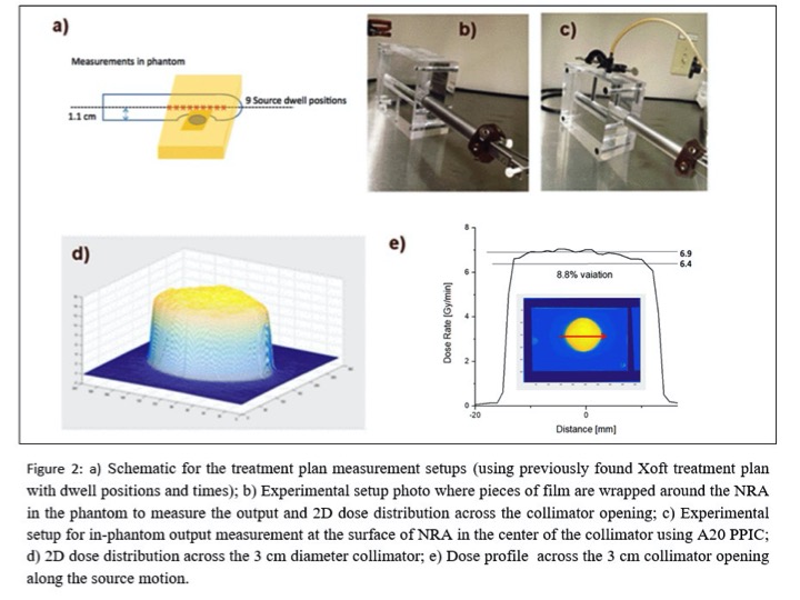
Conclusion
We presented Xoft NRA commissioning process. We described method to measure output and 2D dose distribution using the EBT3 based protocol, and the difference with the PPIC measured output was found to be 8%. When compared to the current applicator, NRA does not have flatting filter and source to surface distance is much shorter (11 vs. 20 mm). Consequently, design of NRA provides, besides the live target monitoring, higher output (6.5 vs. ~2 Gy/min) resulting in significantly shorter treatment times, providing better comfort of patient, and less possibility for movement during treatment.