Quantitative MRI detects radiation dependent demyelination in normal appearing white matter
PO-2081
Abstract
Quantitative MRI detects radiation dependent demyelination in normal appearing white matter
Authors: Anna Ljusberg1,2, Ida Blystad3,2, Peter Lundberg1,2, Emelie Adolfsson1, Anders Tisell1,2
1Department of Medical Radiation Physics, and Department of Health, Medicine and Caring Sciences, Linköping University, Linköping, Sweden; 2Center for Medical Image Science and Visualization (CMIV), Linköping University, Linköping, Sweden; 3Department of Radiology in Linköping, and Department of Health, Medicine and Caring Sciences, Linköping University, Linköping, Sweden
Show Affiliations
Hide Affiliations
Purpose or Objective
This study is using quantitative MRI (qMRI) for longitudinal follow-up after concomitant chemoradiotherapy (CRT) for glioma patients. The aim is to observe changes in normal appearing white matter (NAWM) in the brain and how it is related to the absorbed dose from radiotherapy.
Material and Methods
NAWM were analysed for 10 patients on their follow-up MRI-scans after surgery and treatment according to the Stupp regime (60 Gy/30 fx). Patients were examined with MRI before and after surgery and every 3rd month after completed RT. In median 6 follow-up MRI were performed during the studied period of two years after RT. The qMRI sequence SyMRI MaGiC (GE Medical Systems) was added to standard protocols for brain tumour patients. Synthetic T1 and T2 images were reconstructed as well as relaxation rate maps (R1 and R2). Maps of proton density (PD) and myelin concentration was also calculated, all using the SyMRI software (SyntheticMR AB). ROI were placed in up to 16 anatomical locations for every patient, no ROI was drawn if tumour or oedema was present. MRI and dose distribution from Eclipse (Varian) were rigid registered. ROI drawing and image registration was done in MICE toolkit (NONPI Medical AB). Changes in ROI between follow-up and pre operative MR were calculated using Matlab (The MathWorks Inc.). Parameters observed were relaxation rates R1 and R2, PD and the concentration of myelin. Analyses were done in dependence on radiation dose from CRT and time after treatment.
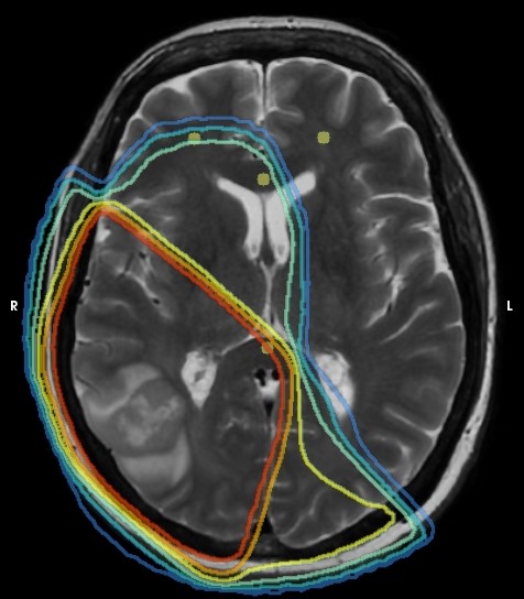
Fig. 1: Synthetic T2-weighted image with ROI (yellow circles) placed bilateral in the lower frontal lobe and in corpus callosum. Isodose levels corresponding to 10, 20, 30, 40, 50 and 57 (95%) Gy.
Results
A significant increase in PD (1.5%) between pre-operative imaging and 1st follow-up, and a corresponding decrease in myelin concentration (-2.2%) were observed. There was no significant change in R1, although a significant change (0.3 s-1) was observed in R2. In Fig. 2 the mean of difference for ROIs with different dose levels is shown as a function of time. With time and low dose, the PD-value and myelin concentration returned to baseline, while for high doses (>30 Gy) the change increased with time.
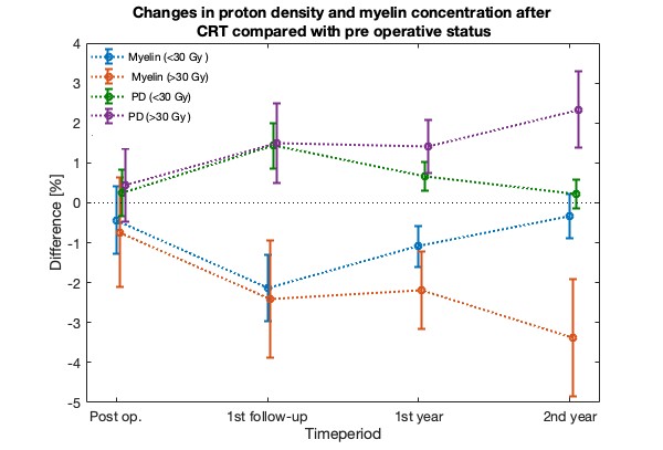
Fig. 2: Mean of differences in ROI for proton density and myelin concentration at different time points. Mean value for ROI at two different dose levels are shown, below or above 30 Gy.
Conclusion
The changes can be interpreted as a demyelination dependent on absorbed dose. For low doses, but not for high doses, myelin was repaired with time. The post-operative values showed that the changes did not depend on surgery but as a consequence of CRT only.
In conclusion, qMRI is a powerful tool, that can be used in a clinical setting, for detecting radiation induced demyelination in NAWM in glioma patients. In the future this knowledge may contribute to an improved radiation therapy planning thereby improving clinical outcome.