Oxygen sensitivity of a silicone based magnetic resonance contrast agent
PO-2073
Abstract
Oxygen sensitivity of a silicone based magnetic resonance contrast agent
Authors: Robert Cormack1, Junichi Tokuda2, Gregory Ekchian3, Evangelina Kaza4, Michael Dyer1, Michael Cima5
1Brigham and Women's Hospital, Radiation Oncology, Boston, USA; 2Brigham and Women's Hospital, Radiology, Boston, USA; 3StratagenBio, -, Cambridge, USA; 4Brigham and Women's Hospital, Radiation Oncolgy, Boston, USA; 5MIT, Koch Institute, Cambridge, USA
Show Affiliations
Hide Affiliations
Purpose or Objective
Hypoxic tumor cells are known to be less radiosensitive than well oxygenated cells, and hypoxia is a poor prognostic factor in prostate, cervical and head and neck solid tumors. Knowledge about tumor oxygen environment could allow radiation dose distributions to be planned with higher doses in the hypoxic regions to compensate for their relative radio resistance. A silicone based material has an MR signal that correlates to blood oxygen content in animal models. The material is biocompatible and, in a solid form, could be implanted in to measure oxygen levels at multiple locations in tumors or monitor oxygen levels over long term follow up. Such sensors could be deployed in brachytherapy procedures or as tumor fiducials in external beam radiation as part of current standard of care procedures. This work assesses the sensitivity of the MR T1 relaxation rate to oxygen levels in a solid form of the material.
Material and Methods
Solid formulations of silicone material were placed in chambers to which a known mixture of gas could be supplied. The chambers were embedded in phantoms containing a solid paraffin phase change material (PCM) having a transition temperature of 37 C to ensure a constant temperature over the duration of a measurement (Figure 1). The phantoms were heated to the PCM’s melting point in a water bath while maintaining a steady flow of gas of known mixture of oxygen and nitrogen. The phantoms were transferred to an MR scanner for measurement of the T1 relaxation rate of the silicone material while continuing to maintain gas flow. The silicone material was scanned in a 3 tesla MR scanner. The phantoms were scanned with inversion recovery sequence for inversion times ranging from 200 to 2000 mS. The T1 relaxation rate of the material was determined from a 3 parameter exponential fit. Multiple experiments were performed varying the oxygen content from 0 to 50% to measure the oxygen dependence of the relaxation rate. 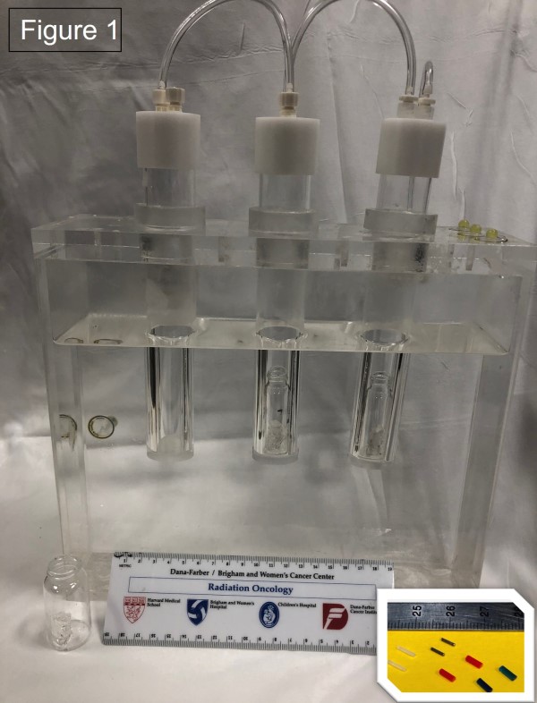
Results
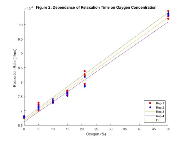
The T1 relaxation rate was fit to a line (Figure 2). The T1 relaxation rate of the material in a 21% oxygen environment at 37C extracted from the fit was 8.17x10⁻⁴ [8.08-8.28x10⁻⁴] mS-1. With a slope of 7.19x10⁻⁶ [6.96-7.41x10⁻⁶] mS-1/%02.
Conclusion
The silicone material offers the ability to manufacture passive implantable oxygen sensors that can be read out by MR scans. Deployed in brachytherapy needles or fiducial markers, this material provides the ability to measure the spatial and temporal variation of tumor oxygen levels which could inform adaptive treatment planning and monitoring patient response. Future work will determine the optimal material composition to achieve the needed SNR for measurements while maintaining the physical characteristics needed for an implantable device. Implantable MR based oxygen sensors (Figure 1 insert, blue and red silicone compared to brachytherapy seeds and spacers) may provide a means to customize radiation dose plans based on measurements of tumor hypoxia.