Vienna, Austria

ESTRO 2023

Session Item

Physical aspects of quantitative functional and biological imaging
7010
Poster (Digital)
Physics
Optimising the MRL Consortium Biomarkers Group Prostate DWI Sequence Image Quality & ADC Comparison
Christopher Moore, United Kingdom
PO-2075

Abstract

Optimising the MRL Consortium Biomarkers Group Prostate DWI Sequence Image Quality & ADC Comparison
Authors:

Christopher Moore1, Abigael Clough2, Claire Nelder2, Cynthia Eccles2, Peter Hoskin3, Rob Chuter1, Jim Zhong3, Damien McHugh1, Ananya Choudhury4, Michael Dubec1

1The Christie NHS Foundation Trust, CMPE, Manchester, United Kingdom; 2The Christie NHS Foundation Trust, Radiotherapy Services, Manchester, United Kingdom; 3The Christie NHS Foundation Trust, Clinical Oncology, Manchester, United Kingdom; 4The Christie NHS Foundation Trust, Research and Innovation, Manchester, United Kingdom

Show Affiliations
Purpose or Objective

The MR Linac Consortium biomarkers working group Prostate DWI sequence was used to measure median prostate ADC in the prostate of patients undergoing treatment on the MR Linac (MRL). The sequence suffered from poor SNR and resolution which limited clinical value. The aim of this work was to improve image quality without compromising the quantitative utility of the sequence and maintaining a reasonable acquisition time.

Material and Methods

The consortium sequence (SS-EPI DWI, b = 0, 150, 500 s/mm2 (2, 4, 16 averages respectively), TE/TR = 82/3354 ms, voxel = 1.9x1.9x4.0 mm, Averages = 1, TA = 3:31) was modified to improve in-plane resolution and increase SNR (SS-EPI DWI, b = 150, 500 s/mm2 (5, 12 averages respectively), TE/TR = 70/2800 ms, voxel = 1.4x1.4x5.0 mm2, Averages = 2, TA = 4:48). The b = 0 image was removed as it is not used to calculate the ADC map. Additional averages were added to the b = 150 and 500 s/mm2 images to improve SNR.

Thirteen patients treated on the MRL, recruited to the MOMENTUM study, were scanned with both sequences at their MR planning session.

Visual image quality assessment of the DWI images and ADC maps was undertaken by a radiologist who assessed the appearance of the whole prostate, transitional/peripheral zones (TZ/PZ), tumour and seminal vesicles against the MOMENTUM image quality assessment criteria (1 = not scanned, 2 = not visible, 3 = not clear, 4 = clear, 5 = very clear), while blinded to the sequence used.

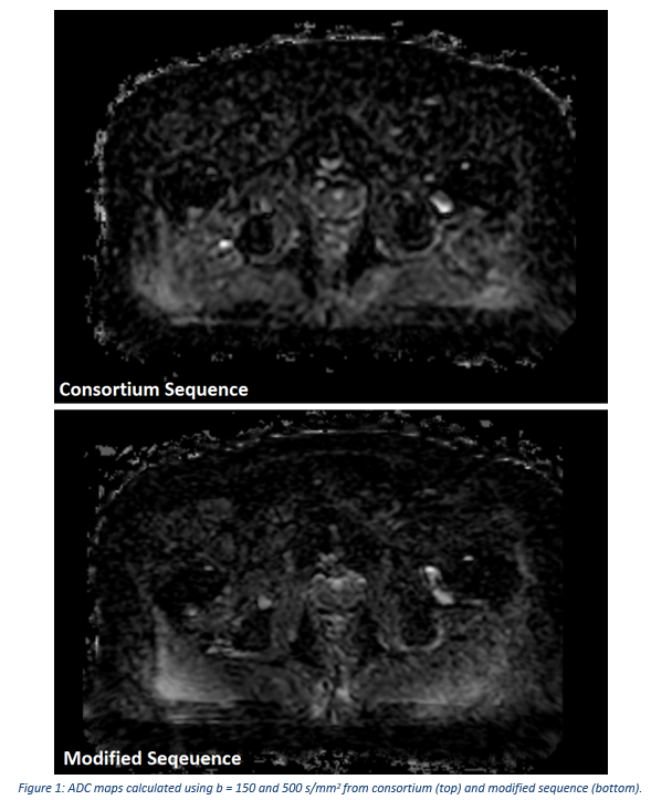
ADC maps (figure 1) were calculated using the b = 150 and 500 s/mm2 images. Structure contours were obtained by rigidly registering the DWI images to the planning MR images, and adapting the contours drawn on the planning MR. Median whole prostate ADC measurements were compared between the two sequences by Bland-Altmann analysis.


Results

Figure 2a shows the number of times each sequence was scored higher for each structure examined on the DWI and ADC images.

The Bland-Altman analysis (Figure 2b) shows the results of the quantitative comparison between the two sequences.


Conclusion

The modified sequence performed as well as or better than the consortium sequence, in the qualitative image quality assessment for 92% (DWI & ADC) of comparisons for whole prostate, peripheral/transitional zone and tumour visualisation. The modified sequence performed as well as or better that the consortium sequence for 77% (DWI) /69% (ADC) of comparisons for seminal vesicle visualisation. These results indicate that the modified sequence is comparable to, or improves, images quality.

In the comparison of ADC, the modified sequence measured median prostate ADC on average 5% lower than the consortium sequence, indicating a systematic difference between the two sequences. ADC measurements from the modified sequence should be directly compared to those obtained from the consortium sequence.

Further work will be undertaken to use the modified sequence to measure ADC throughout treatment to detect ADC changes.