Voxel-wise tumour response estimation from functional imaging and radiobiological model.
PO-2070
Abstract
Voxel-wise tumour response estimation from functional imaging and radiobiological model.
Authors: Antonio Lopez Medina1, Manuel Porta-Lorenzo2, Mercedes Riveira-Martin3, Isaac Sanchez-Diaz4, Ricardo Dorado-Dorado1, Francisco Javier Salvador-Gomez1, Íñigo Nieto-Regueira5, Virginia Ochagavia-Galilea5, Víctor Muñoz-Garzón5
1Hospital do Meixoeiro - Galaria - SERGAS, Radiofisica y PR, Vigo, Spain; 2University of Vigo, atlanTTic Research Center, Vigo, Spain; 3Instituto de Investigación Sanitaria Galicia Sur , Medical Physics, Vigo, Spain; 4Instituto de Investigación Sanitaria Galicia Sur, Medical Physics, Vigo, Spain; 5Hospital do Meixoeiro - Galaria - SERGAS, Radioterapia, Vigo, Spain
Show Affiliations
Hide Affiliations
Purpose or Objective
To implement biologically guided radiotherapy, multiple tools must be developed to utilise the information contained in functional images. We have developed a script integrated in one of the most widely used TPS to predict the evolution of tumour cell density using a radiobiological model that includes information from functional images, such as diffusion-weighted imaging (DWI) and dynamic contrast-enhanced imaging (DCE).
Material and Methods
An Eclipse Scripting Application Programming Interface (ESAPI) script of ARIA (Varian Medical Systems, version 15.1) has been developed in the C# programming language for individualised calculation of tumour response. For a selected patient, the program can work with data from ARIA (image datasets, plans, dose distributions, contours…) or any DICOM file. In the case of importing new DICOM images, they must be previously registered to an image set registered to the planning CT in ARIA.
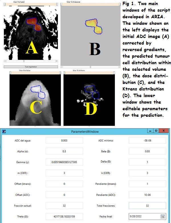
The programme obtains a voxel-wise estimate of the tumour cell density and the apparent diffusion coefficient (ADC) maps, seizing the correlation between ADC values and cellularity [doi.org/10.1038/s41598-021-87887-4]. The input data required are an initial ADC map, obtained from a low-distorted DWI sequence, a DCE Ktrans map, the dose distribution and the treatment schedule. The cell response was calculated based on the linear quadratic model (LQ) modified by oxygenation and proliferation. Parameters (Fig. 1) can be configured individually. We considered that vascularisation is proportional to the value of Ktrans and that oxygenation can be modelled as a function of Ktrans. Optionally, several ADC maps corresponding to different stages of the treatment can be loaded to compare the estimated tumour cell density from ADC maps and from radiobiological model.

The parameters of the LQ model were obtained from the literature [doi.org/10.1088/0031-9155/53/17/001], but they can be edited to fit the available data. The interpolation method used is nearest neighbour.
Results
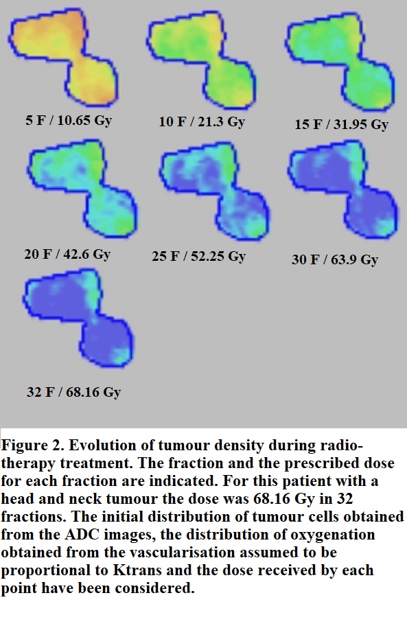
We show an example performed on a patient recruited for the ARTFIbio project treated with 68.16 Gy in 32 fractions (Fig. 2). The initial ADC map was calculated with b = 0, 1000 and distortion corrected by reversed-gradient method.

The ADC for different stages and the tumour cell density within the treatment was calculated with the script. As seen in Fig. 2 the hypoxic areas (poorly vascularised areas) of the tumour show lower response to the treatment at all the fractions.
The inhomogeneous response of the tumour due to the effects of poor oxygenation can be observed. The use of such tools can be very useful in order to design a voxel-wise personalised treatment.
Conclusion
We have developed a software in a widely used environment (Eclipse-ARIA), which allows the integration of relevant biological information in functional images for the prediction of tumour response in a routine workflow.
SINFONIA project funded from the Euratom research and training programme 2019-2020 under grant agreement No 945196