Hounsfield units to electron density conversion: inhomogenous phantoms, user experience and RTP dose
PO-1767
Abstract
Hounsfield units to electron density conversion: inhomogenous phantoms, user experience and RTP dose
Authors: Wolfgang Baus1, Georg Altenstein2, Horst Hermani1, Christos Moustakis3
1Robert Janker Klinik, Radiotherapy and Radiooncology, Bonn, Germany; 2University Hospital of Cologne, Radiooncology, Cologne, Germany; 3University Hospital Muenster, Radiotherapy and Radiooncology, Münster, Germany
Show Affiliations
Hide Affiliations
Purpose or Objective
Conversion of computed tomography (CT) Hounsfield Units (HU), to electron density (ED) or material via lookup-table (LUT) is the basis of heterogeneity corrected dose calculation for radiotherapy treatment planning (RTP). Though the necessary precision of the conversion might be debatable [1], using an ED-phantom at commissioning of the RTP-System (RTPS) is standard [2]. However, CT measures mainly absorption by photo effect (depending on effective atomic number, Zeff), whereas dose in radiotherapy is caused rather by Compton scatter (depending on ED). Neglecting this fact by using unsuitable phantom material can lead to an inappropriate LUT (iLUT) as was observed for one of our RTPS. Therefore we investigated phantoms and materials. In addition, the LUTs used by the participants of an RTP study [3] are compared and the clinical impact is investigated.
Material and Methods
We measured 3 commercial (CIRS 062, RMI 467, Catphan 504) and self-built phantoms (SBP) with CT-scanners from 3 vendors using various scan parameters. The SBP contained, CaCO3, hydroxylapatite, ethanol, n-propane, aqueous solutions (aqus.) of CaCl2, and water. For theoretical understanding, Zeff of various compounds was calculated after modifying a python script gratefully provided by Liu ([4]). To estimate the influence of the iLUT, patient plans covering bony anatomy were calculated and compared. In addition, plans using different LUTs where compared investigating the influence on plan metrics as part of a multicentre study on pancreas planning (PACA).
Results
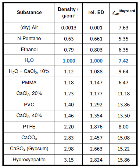
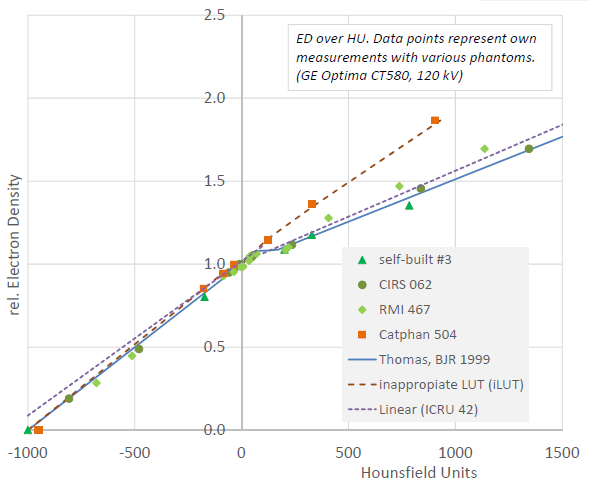
Fig. 1 shows a graph of ED over HU for different materials, including iLUT and standard curves ([1], [5]). Nearly all data points lie in the vicinity of the standard curves – except the ones of the Catphan-phantom. These lie on a straight line through the origin since Zeff of its inhomogeneities (e.g. PTFE, see Tab. 1) are near water. Though the iLUT is markedly different, the resulting dose difference was only between 0.7 and 1.3 % (head, pelvis). For PACA, different LUTs resulted in changes of plan metrics of generally less than 1.0 %, though in some cases it could be up to 1.3 % (median PTV dose) for targets, and for organs at risk 1.5 % in high dose regions and several % in low dose regions.
Conclusion
Inhomogeneity phantoms for RTP should comprise both water- and bone-like materials, Zeff being the decisive parameter. As bone surrogate aqus. of CaCl2 seem well suitable. PTFE and other materials with water-like Zeff, though included in commercial phantoms, are definitely not suitable as bone surrogate. However, for photon plans the conversion LUT has rather limited clinical relevance, especially compared to changes in tube voltage, and measurements should always stand the comparison to standard values.
Literature
[1] Thomas, Br J Radiol (1999)
[2] Smilowitz et al., JACMP (2015)
[3] PACA-Study, to be published
[4] Liu et al., Med Phys (2021)
[5] ICRU Report 42, Bethesda (1987)

Tab. 1 Phantom materials

Fig. 1 Hounsfield to electron density conversion