Deep-learning standardization of MR images in radiomic studies-Application to LACC T2w MRI
Stephane Niyoteka,
France
PO-2093
Abstract
Deep-learning standardization of MR images in radiomic studies-Application to LACC T2w MRI
Authors: Stephane Niyoteka1, Rahimeh Rouhi1, Pierre-Antoine Laurent1, Alexandre Carré1, Samir Achkar1, Sebastien Diffetocq2, Corinne Balleyguier2, Cyrus Chargari1, Eric Deutsch1, Charlotte Robert1
1Gustave Roussy Cancer Campus, Radiation Oncology, Villejuif, France; 2Gustave Roussy Cancer Campus, Radiology, Villejuif, France
Show Affiliations
Hide Affiliations
Purpose or Objective
Radiomic features are highly dependent on image acquisition and reconstruction parameters especially in MR imaging. Many methods are available to standardize MR images but no recommendation exists to date regarding the optimal method for radiomic analyses of the pelvic region. The objective of this study was to propose a generative adversarial network-based method to solve this problem by considering images of locally advanced cervical cancer (LACC) patients acquired for several parameter sets, and to compare it with image post-processing and the Combat method.
Material and Methods
T2-weighted (T2w) MR images from 30 patients treated for LACC were acquired prospectively. For each patient, T2w MR images were taken sequentially using a 1.5T GE Optima MR450w (GE Healthcare, Waukesha, WI) considering modifications in repetition time (TR) and voxel (VS): 1) TR>5700ms and VS=0.58×0.58×4.0mm3, 2) TR = 3300ms and VS=0.78x0.78×4.0mm3, 3) 3rd acquisition with TR=3300ms and VS=0.58×0.58×4.0mm3. On each image, the GTV was manually segmented. Bias field correction and voxel resampling were applied to all images. On one side, MR images were standardized using either a histogram-matching strategy (Nyul) or z-score normalization. Radiomic features were then extracted with Pyradiomics. On the other hand, the ComBat harmonization method was applied to features extracted from native MRI corrected from the bias field and resampled. We also applied the ComBat harmonization method to radiomic features extracted after Nyul or z-score image standardization. Lastly, a GAN was trained using 40 paired images from the 1st and 2nd acquisitions to generate MR images standardized to the 3rd acquisition (20 patients). The trained GAN was then tested on the remaining 10 patients. By means of Intra-Class Correlation (ICC) and Concordance Correlation Coefficient (CCC), robust features were assessed (CCC & ICC >0.75) for each standardization method.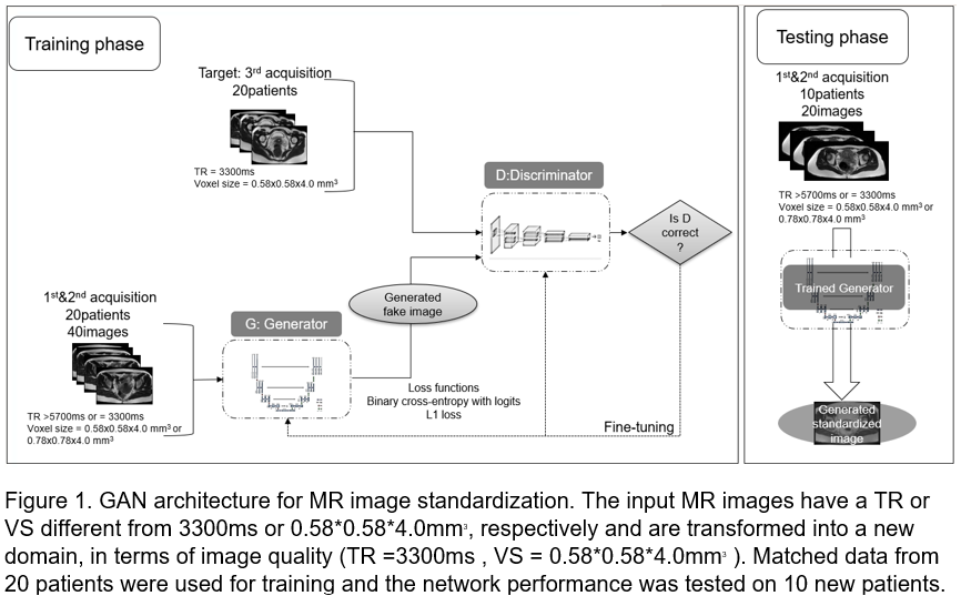
Results
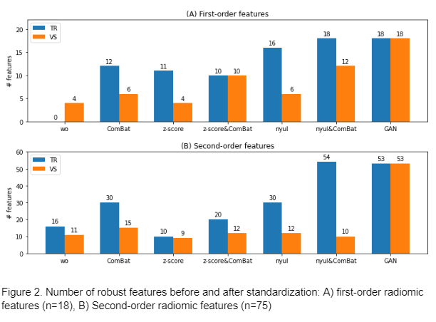
We found that none of 1st order features was robust to TR modulation and only 22% were robust to VS changes. For 1st order features, Nyul followed by ComBat increased the features robust to TR and VS changes to 67% and 100% respectively. In the test set, GAN standardization resulted in 100% robust 1st order features for both TR and VS modulation.
For second-order features, the histogram matching technique combined with ComBat led to 20% and 72% features robust to TR and VS shifts, respectively, while GAN normalization resulted in 71% features robust to TR and VS modulation
 .
.
Conclusion
Despite the encouraging results of combining image normalization and ComBat, this technique is still complex to implement in a multicenter setting. Our results suggest that deep learning-based methods could provide a tool capable of simultaneously mitigating the effects of multiple acquisition/reconstruction parameters on radiomic features. A normalization method based on a Cycle-GAN will be implemented on multi-scanner MR images and its normalization power will be evaluated.