Vienna, Austria

ESTRO 2023

Session Item

Upper GI
6010
Poster (Digital)
Clinical
Comparison of Lung Doses and Dependent Factors in IMRT, Tomotherapy & 3DCRT Planning of Ca Esophagus
Manasa Penumur, India
PO-1345

Abstract

Comparison of Lung Doses and Dependent Factors in IMRT, Tomotherapy & 3DCRT Planning of Ca Esophagus
Authors:

Manasa Penumur1, Smriti Goswami1, Bibhash Chandra Goswami1, Diplu Choudhury1, Fahim Arman Hassan1, Ghritashee Bora1, Moumita Paul1, Luri Borah1, Rheetwik Barauh1, Dimpal Saikia1

1State Cancer Institute, Gauhati Medical College, Radiation Oncology, Guwahati, India

Show Affiliations
Purpose or Objective

Definitive chemoradiation is the treatment of choice for surgically or medically inoperable patients of esophageal cancer. Intensity Modulated Radiotherapy (IMRT) and Helical Tomotherapy (HT) planning techniques provide better conformality of dose distribution and sparing of organs at risk when compared to Three-Dimensional Conformal Radiotherapy (3DCRT), but with the risk of increased low dose bath.

Material and Methods

A total of 28 patients of carcinoma esophagus planned for definitive chemoradiation were recruited from June 2021 till September 2022. Patients were planned for a total dose of 50.4Gy. All patients underwent CT simulation with intravenous contrast, and the treatment volumes and organs at risk were contoured according to RTOG contouring guidelines. Treatment plans of IMRT, HT and 3DCRT were generated for each patient. The lung dose parameters of V20, V30, V5, V10 and Dmean were noted for each plan. Data on location of GTV, volume of GTV and PTV, and craniocaudal length of PTV were the noted factors. The data was compared and analyzed using SPSS software version 28.0.1.0 with Friedman test and Pearson’s correlation test, and a p value of 0.05 was considered as significant.

Results

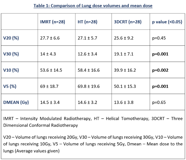
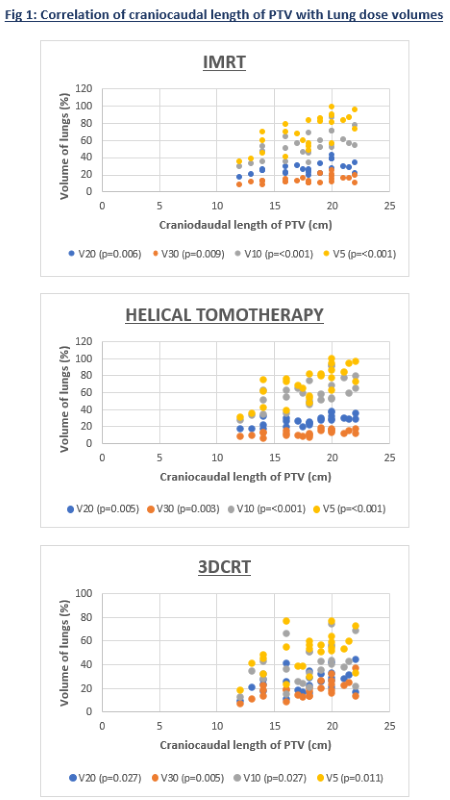
The Dmean and V20 lung volumes were similar between IMRT, HT and 3DCRT (Table 1). IMRT and HT had significantly lower V30 when compared to 3DCRT (14 ± 4.3, 12.6 ± 3.4, 19.1 ± 7.1 respectively; p=0.001), whereas V5 (69 ± 18.7, 69.8 ± 19.6, 50.1 ± 15.3 respectively; p=0.001) and V10 (53.6 ± 14.5, 58.4 ± 16.6, 39.9 ± 16.2 respectively; p=0.002) were significantly lower in 3DCRT. All lung dose volumes had significant moderately positive to strongly positive correlation with craniocaudal length of the PTV across the three different techniques (Figure 1). Volume of PTV had no significant correlation with lung dose volumes, whereas volume of GTV had moderately positive correlation with V20 of IMRT (Correlation Coefficient Factor(R)=0.43; p=0.02). IMRT had significantly less low dose region in upper third esophageal tumors (GTV - Gross Tumor Volume) compared to mid and lower thoracic location in V5 (55.1 ± 15.7, 80.2 ± 14.8, 70.2 ± 12.3 respectively; p=0.016), V10 (44.3 ± 12.4, 60.4 ± 13.2, 57.3 ± 11.5 respectively; p=0.017), and Dmean (12.3 ± 3, 16.3 ± 3.3, 14.7 ± 1.1 respectively; p=0.015). HT also had lower V5 (55.5 ± 16.7, 81.3 ± 16.1, 71.8 ± 9.1 respectively; p=0.011), V10 (47.7 ± 12.7, 65.3 ± 17, 66.4 ± 7 respectively; p=0.042), and Dmean (12.5 ± 2.5, 16.5 ± 3.2, 16.3 ± 1 respectively, p=0.01) in upper third esophageal tumors.


Conclusion

IMRT and HT provide similar dose constraints achieved to the lungs when compared to 3DCRT planning but with a significant increase in the low dose volume. Higher dose volumes are observed in increasing length of PTV in all three techniques. Upper third esophageal lesions in IMRT and HT have lesser low dose irradiation. However, randomized studies with a larger sample size and follow up of the patients are required to identify long term clinical effects and outcomes.