Use of OSLDs for dosimetric verification of Helical TomoTherapy dynamic field width treatment plans
Nikolaos Prountzos,
Greece
PO-1623
Abstract
Use of OSLDs for dosimetric verification of Helical TomoTherapy dynamic field width treatment plans
Authors: Nikolaos Prountzos1, Evaggelos Pantelis2, Panagiotis Papagiannis1, Pantelis Karaiskos1, Argyris Moutsatsos3, Eleftherios Pappas3, Nikolaos Fotos4, Stamatina Kanellopoulou5
1National and Kapodistrian University of Athens, Medical School, Medical Physics Laboratory, Athens, Greece; 2National and Kapodistrian University of Athens, Medical School, Medical Physics Laboratory, Athens, Greece; 3Iatropolis Clinic, Radiotherapy Department, Athens, Greece; 4MediRay Inc, Dosimetry Laboratory, Athens, Greece; 5MediRay Inc, Dosimetry Laboratory , Athens, Greece
Show Affiliations
Hide Affiliations
Purpose or Objective
To evaluate the use
of Optically Stimulated Luminescence Dosimeters (OSLDs) for the verification of
Helical TomoTherapy (HT) (Accuray Inc, CA, USA) treatment plans using the dynamic
jaw delivery feature.
Material and Methods
A batch of nanoDot OSLDs
(Landauer Inc, IL, USA) were used. The dosimeters were calibrated in terms of
dose to water using a 6MV x-ray photon beam for doses up to 300cGy. Due to the
helical treatment delivery fashion, radiation is incident to the dosimeters from
variable directions. Therefore, Monte Carlo (MC) simulations were performed to assess
potential directional dependence of nanoDot response using the EGSnrc MC code
and the egs_chamber user code. To model the dosimeters, the C++ geometrical
package available with the EGSnrc and information found in the literature and
vendor manuals were used. For the measurement setup, RW3 (PTW, Freiburg,
Germany) slabs of 13cm total thickness were used. The central slab was appropriately
machined to hold the OSLDs in axial and coronal orientations. The phantom was
CT scanned and two HT treatment plans were developed using the dynamic jaw
delivery option of the HT systems. The first plan involved the irradiation of 5
targets situated along the central craniocaudal axis of the slab-phantom
delivering 2Gy. In the second plan, 3 targets centered on the phantom
craniocaudal axis lying across the Left-Right direction were irradiated
delivering 2Gy on the side targets while a boost dose of 2.5Gy was planned for
a sub-volume of the central target mimicking the delivery of simultaneously
integrated boost (SIB). NanoDot measurements were performed at dose-plateau areas
(plans 1&2), as well as regions of dynamic jaw movement (plan 1). Phantom
alignment at irradiation position was performed using the image guidance capabilities
of the HT system.
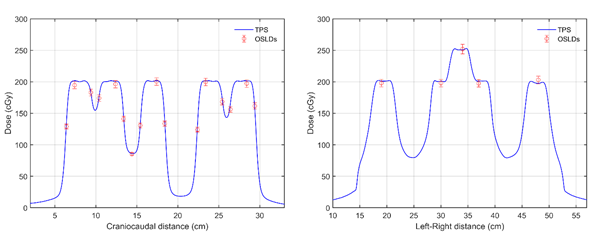
Results
MC simulations
revealed mirror directional dependence of nanodot response, being up to 5% when
irradiated from an angle of 90o. Experimental dosimetry results are
presented in figure 1 along with corresponding profile data exported from the
PrecisionTM treatment planning system (TPS). As seen, nanoDot
measurements agree with TPS predictions within experimental uncertainties of 5%.
Conclusion
Exhibiting minimal directional
dose-response dependence, in agreement with the corresponding literature, the nanoDots
can provide precise dosimetry measurements allowing for the experimental verification
of HT treatment plans.