ESTRO 2024 Congress report
Madelon van den Dobbelsteen, Pim T.S. Borman, Laurie J.M. de Vries, Sara L. Hackett, Kalin I. Penev, Rocco Flores, Stephanie Smith, Yoan LeChasseur, Simon Lambert-Girard, Benjamin Côté, Peter L. Woodhead, Lando S. Bosma, Cornel Zachiu, Bas W. Raaymakers, Martin F. Fast
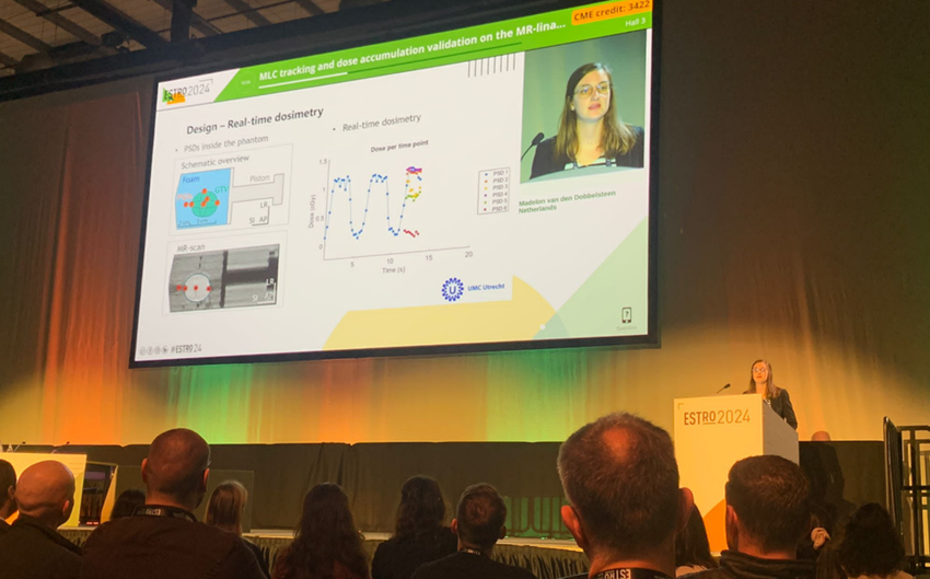
Study motivation: We can acquire online MRI scans on the MR-linac that capture the full complexity of anatomical motion during delivery of treatment. This time-resolved anatomical information is increasingly used to facilitate novel dose accumulation and (real-time) plan adaptation techniques. During the ESTRO conference, several sessions were dedicated to dose accumulation and real-time motion-management techniques. Challenges await us regarding the introduction of these methods into the clinic. Some steps remain to be taken to bridge the bench-to-bedside gap and to take these initiatives into a clinical setting. One of the essential components is a reliable quality assurance tool for verification. Therefore, we developed an MRI-compatible prototype (which was designed in collaboration with IBA QUASAR, Medscint and University Medical Center Utrecht) of a deformable motion phantom with integrated plastic scintillation dosimeters (PSDs). This phantom can be used for end-to-end validation which is an essential step towards the clinical introduction of these newly developed innovations. At ESTRO 2024, we presented the first demonstration of the feasibility of multi-leaf collimator (MLC) tracking and dose accumulation with this phantom during the Intra-fraction motion management and real-time adaptive radiotherapy session (Figure 1).
Most important findings: We used the deformable phantom with six integrated PSDs in a motion phantom to measure the dose in real-time. The tumour model that is embedded in the phantom can achieve translations of 20mm and deformations of 10mm between end-inhale and end-exhale positions. To validate dose accumulation, the statically delivered dose per respiratory phase was warped through the use of three different deformable image registration approaches and compared with a dynamically measured and planned dose. During the MLC tracking experiments, coronal cine MR images were acquired to identify the tumour automatically. Both translational and deformable MLC tracking were tested using sinusoidal motion.
When we focused on the two PSD locations on the edges of the delivered field, which are most affected by motion, the measured dose was found to align with the accumulated dose within positioning uncertainty. As expected, measured and planned doses differed greatly due to the impact of motion, and this underlined the need for active motion management. For one of the PSD locations, the unmitigated motion led to approximately 50% dose loss compared with the planned dose. During MLC tracking experiments, the two PSD locations at the edges of the tumour were strongly affected by sinusoidal motion. The scenario with unmitigated motion showed large dose differences compared with the static scenario, while translational and deformable tracking showed smaller dose differences compared with the static scenario. For one PSD location just outside the target, a maximum dose difference of approximately 5% was shown for the tracking scenarios compared with the dose that was delivered to this PSD location in the static scenario, while unmitigated motion more than doubled the dose.
Research implications: This study demonstrates the vast potential of a novel prototype of a deformable phantom with integrated PSDs for real-time dosimetry measurements on an MR-linac. Currently, we are revising the design and assembly of the phantom to achieve even lower measurement uncertainties. The deformable phantom gives us the opportunity to investigate the latest adaptive radiotherapy innovations. Therefore, we hope that this phantom can help to bridge the care gap for successful end-to-end test validations.

Figure 1: Presentation during the Intra-fraction motion management and real-time adaptive radiotherapy session at ESTRO 2024.
Madelon van den Dobbelsteen

PhD candidate
Department of Radiotherapy
University Medical Center Utrecht
Utrecht, The Netherlands
linkedin.com/in/madelon-van-den-dobbelsteen
m.vandendobbelsteen-3@umcutrecht.nl